. 2022 Dec 1;209(11):2149-2159.
doi: 10.4049/jimmunol.2200324.

Reduced T Cell Priming in Microbially Experienced "Dirty" Mice Results from Limited IL-27 Production by XCR1+ Dendritic Cells

Affiliations

Reduced T Cell Priming in Microbially Experienced "Dirty" Mice Results from Limited IL-27 Production by XCR1+ Dendritic Cells

Frances V Sjaastad et al. J Immunol. .

Abstract

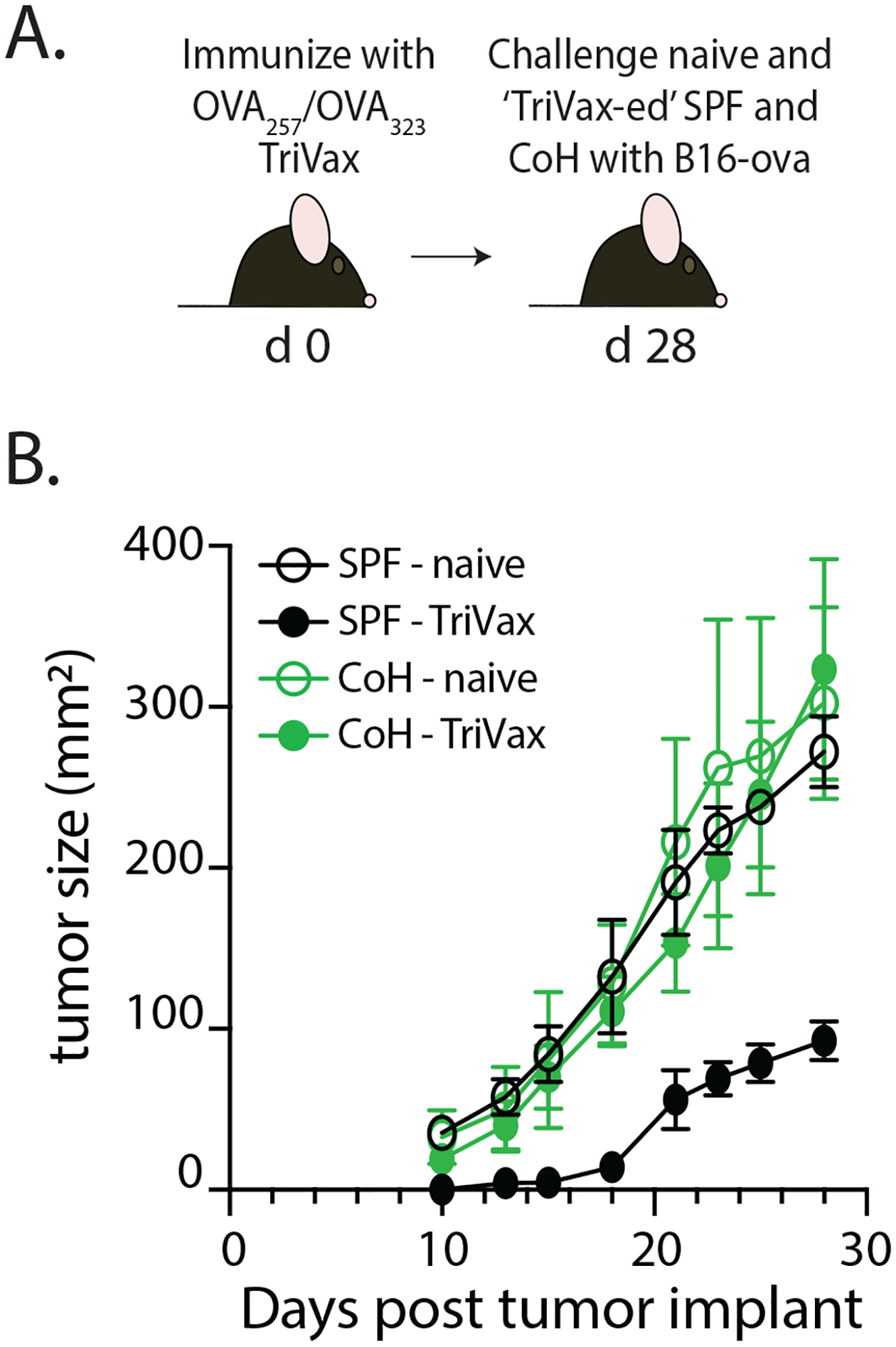
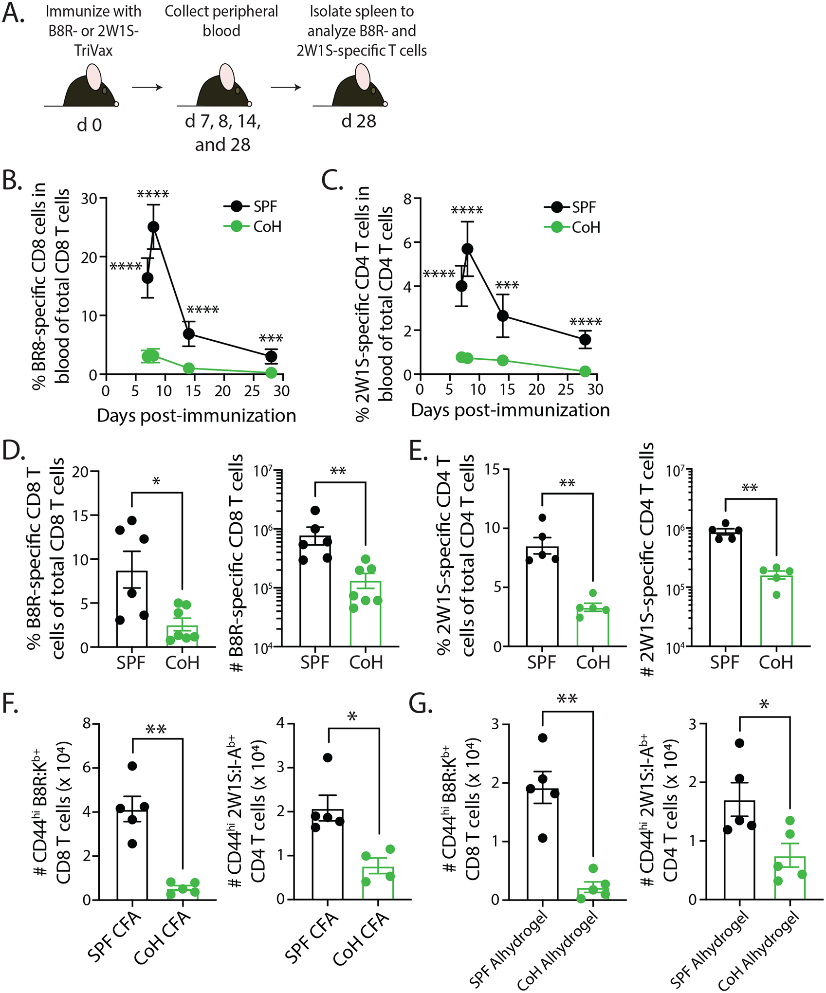
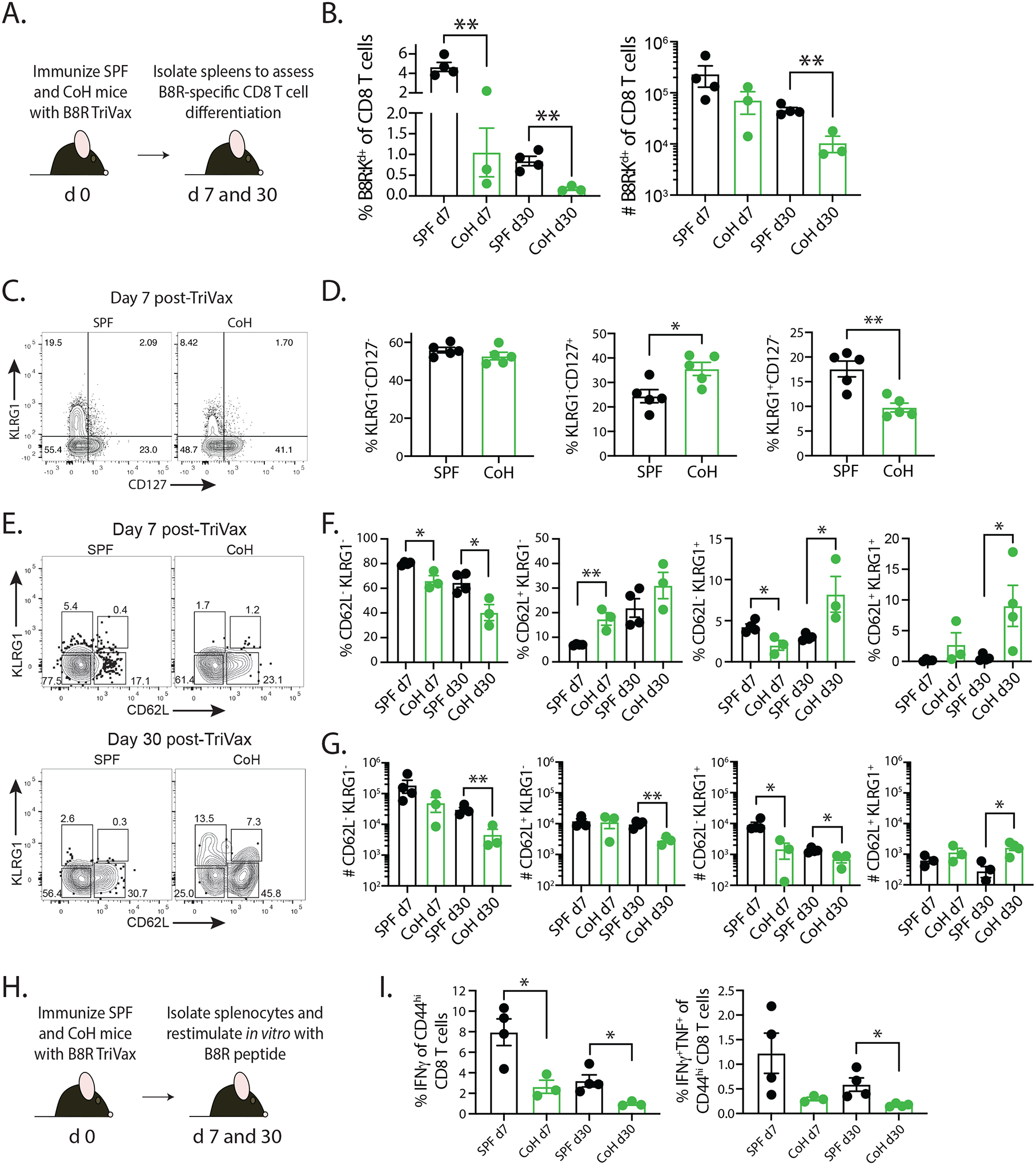
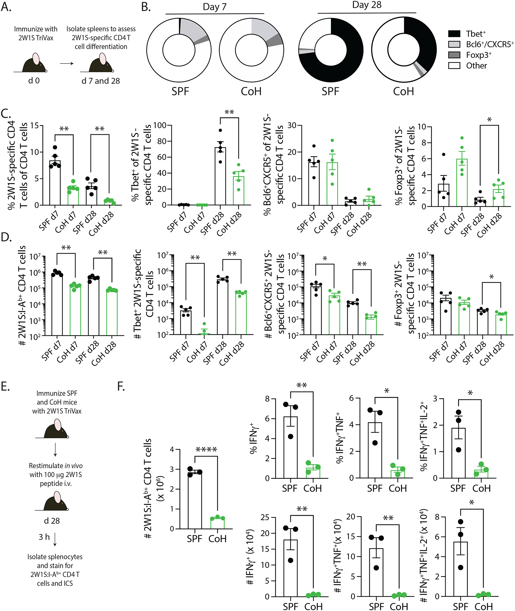
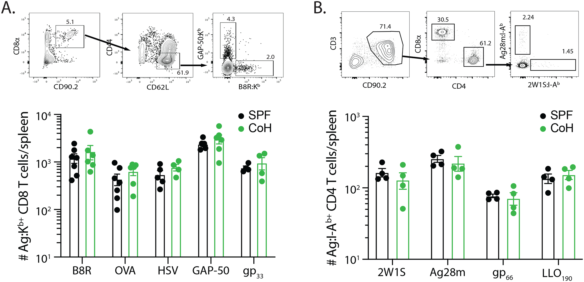
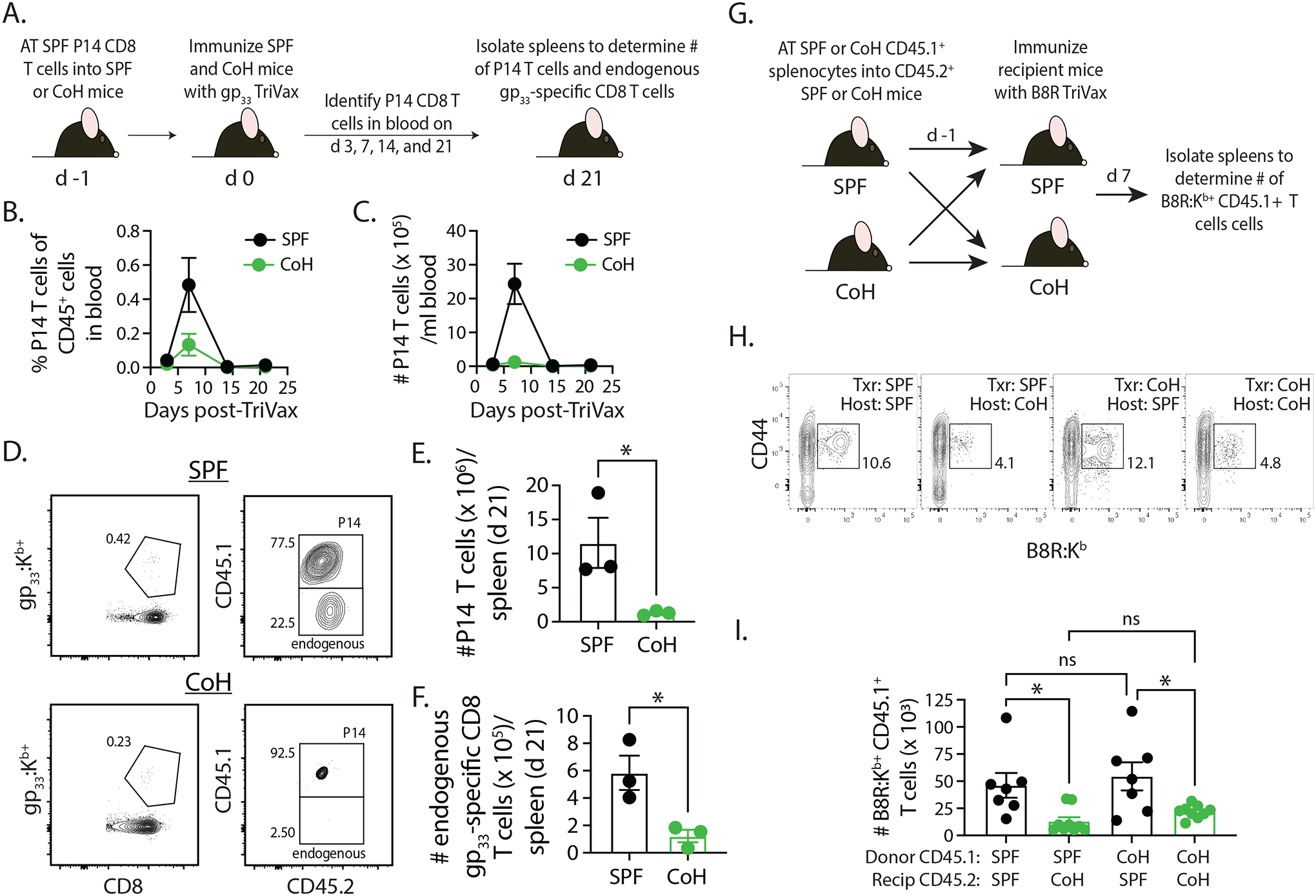
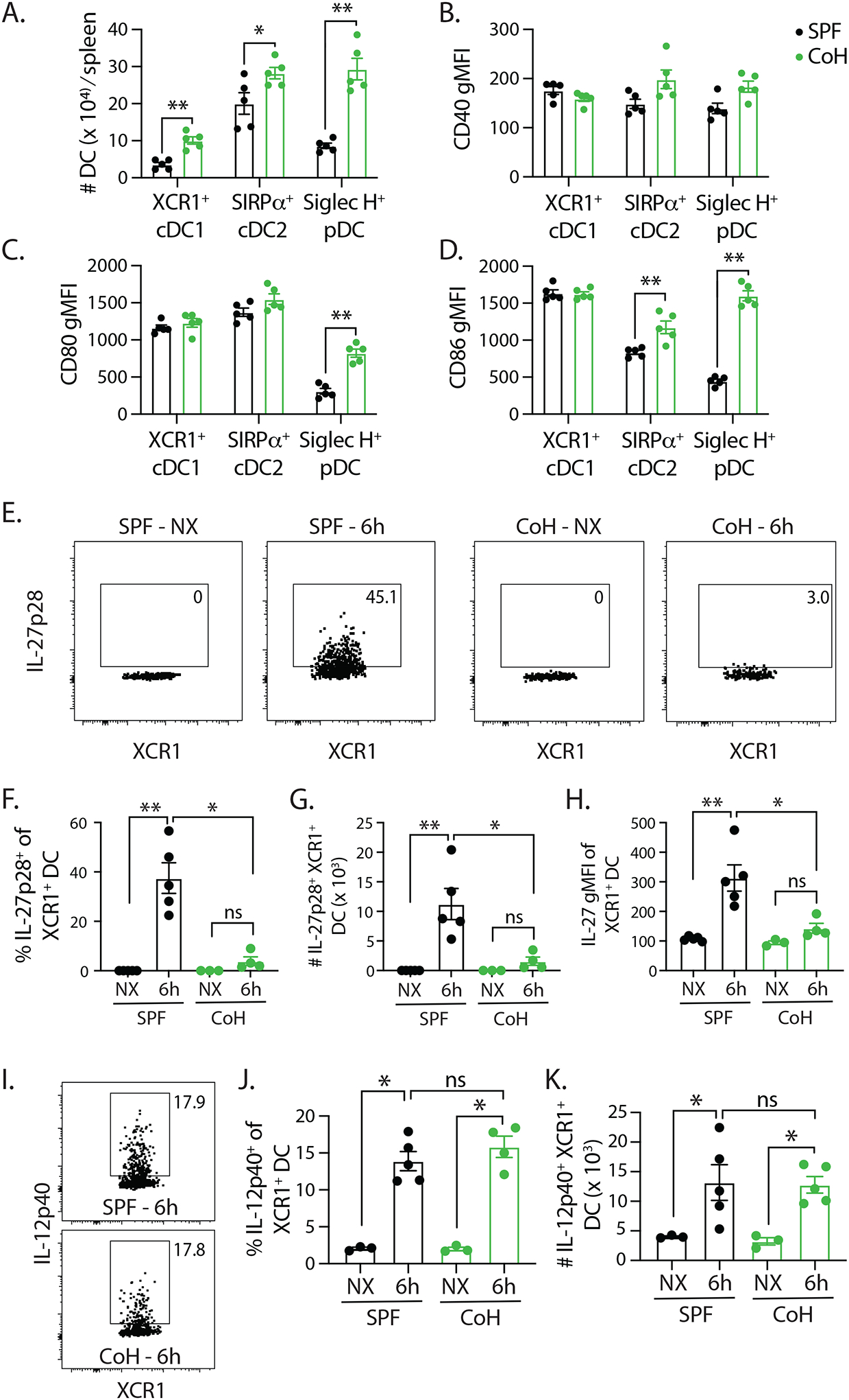
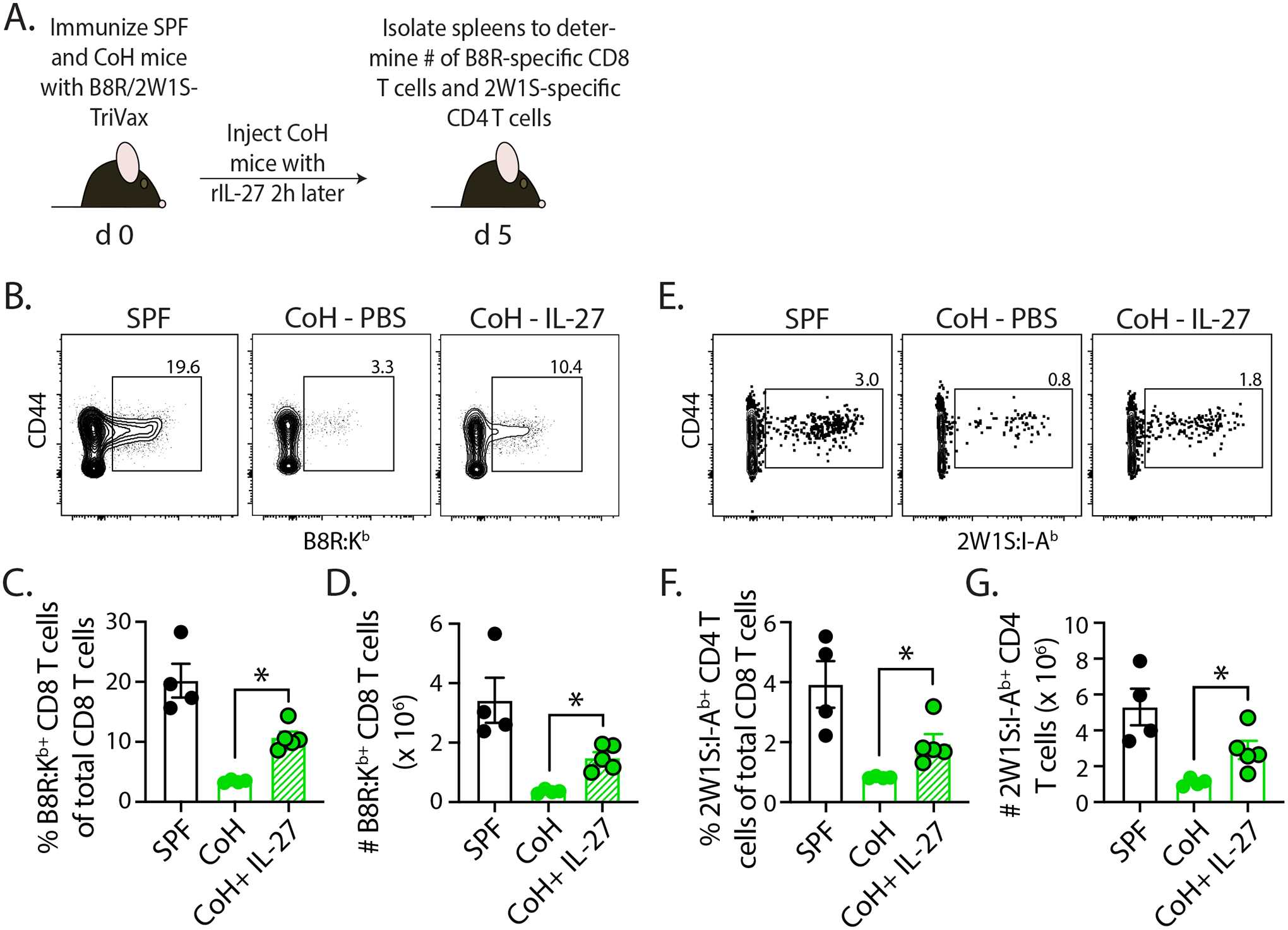
Successful vaccination strategies offer the potential for lifelong immunity against infectious diseases and cancer. There has been increased attention regarding the limited translation of some preclinical findings generated using specific pathogen-free (SPF) laboratory mice to humans. One potential reason for the difference between preclinical and clinical findings lies in maturation status of the immune system at the time of challenge. In this study, we used a "dirty" mouse model, where SPF laboratory mice were cohoused (CoH) with pet store mice to permit microbe transfer and immune system maturation, to investigate the priming of a naive T cell response after vaccination with a peptide subunit mixed with polyinosinic-polycytidylic acid and agonistic anti-CD40 mAb. Although this vaccination platform induced robust antitumor immunity in SPF mice, it failed to do so in microbially experienced CoH mice. Subsequent investigation revealed that despite similar numbers of Ag-specific naive CD4 and CD8 T cell precursors, the expansion, differentiation, and recall responses of these CD4 and CD8 T cell populations in CoH mice were significantly reduced compared with SPF mice after vaccination. Evaluation of the dendritic cell compartment revealed reduced IL-27p28 expression by XCR1+ dendritic cells from CoH mice after vaccination, correlating with reduced T cell expansion. Importantly, administration of recombinant IL-27:EBI3 complex to CoH mice shortly after vaccination significantly boosted Ag-specific CD8 and CD4 T cell expansion, further implicating the defect to be T cell extrinsic. Collectively, our data show the potential limitation of exclusive use of SPF mice when testing vaccine efficacy.


Figures

FIGURE 1.
FIGURE 1.. Reduced antitumor immunity in TriVax-immunized microbially experienced cohoused (CoH) mice.
A. Experimental Design – SPF and CoH B6 mice were immunized with OVA257/OVA323-TriVax. After 28 days, mice were challenged with B16-ova cells (2.5 × 10⁵ cells in 0.1 ml s.c.). B. Tumor growth was then monitored over the next 28 days. As a reference, cohorts of unimmunized SPF and CoH mice were also challenged with B16-ova cells. Data in B are representative of 2 replicate experiments. n = 8 mice/group.
FIGURE 2.
FIGURE 2.. TriVax-immunized CoH mice exhibit blunted CD8 and CD4 T cell expansion.
A. Experimental Design – SPF and CoH B6 mice were immunized with either B8R- or 2W1S-TriVax. Blood was collected on days 7, 8, 14, and 28 after immunization to determine the frequency of (B) B8-specific CD8 T cells or (C) 2W1S-specific CD4 T cells of total CD8 or CD4 T cells, respectively. On day 28, spleens were also isolated to determine the frequency and number of (D) B8R-specific CD8 T cells or (E) 2W1S-specific CD4 T cells. F-G. SPF and CoH mice were immunized with either B8R or 2W1S peptide mixed with either (F) Complete Freund’s Adjuvant (CFA) or (G) Alhydrogel. Spleens were collected 7 days later, and the number of B8R-specific CD8 T cells or 2W1S-specific CD4 T cells were determined. Data in B-G are representative of at least 2 technical replicates. n = 4–7 mice/group. * p ≤ 0.05, ** p ≤ 0.01, *** p ≤ 0.005, **** p ≤ 0.001.
FIGURE 3.
FIGURE 3.. CD8 T cells from TriVax-immunized CoH mice display reduced differentiation and function after restimulation.
A. Experimental design – SPF and CoH B6 mice were immunized with B8R-TriVax. On days 7 and 30 after immunization, spleens were isolated and processed for flow cytometry to determine the (B) frequency and number of B8R-specific CD8 T cells. C-D. Day 7 post-TriVax samples were examined for KLRG1 and CD127 expression to define MPEC (KLRG1CD127+) and SLEC (KLRG1+CD127) frequency. E-G. Day 7 and 30 post-TriVax samples were examined for KLRG1 and CD62L expression to define the frequency and number of KLRG1CD62L (TEM), KLRG1CD62L+ (TCM), and KLRG1+CD62L (LLEC) subsets. Data are representative of at 3 technical replicates consisting of 3–4 mice/group/timepoint. * p ≤ 0.05, ** p ≤ 0.01. H. Experimental design – SPF and CoH B6 mice were immunized with B8R-TriVax. After 7 and 30 days, spleens were collected and processed into a single cell suspension for in vitro restimulation with B8R peptide to induce effector cytokine (IFNγ and TNFα) production. I. The frequency of IFNγ+ and IFNγ+TNFα+ cells among the CD44hi CD8 T cells within the culture was determined by flow cytometry. Data are representative of 2 technical replicates consisting of 3–4 mice/group/timepoint. * p ≤ 0.05
FIGURE 4.
FIGURE 4.. Reduced CD4 T cell differentiation and recall response in TriVax-immunized CoH mice.
A. Experimental design – SPF and CoH B6 mice were immunized with 2W1S-TriVax. On days 7 and 28 after immunization, spleens were isolated and processed for flow cytometry to determine the extent of 2W1S-specific CD4 T cell differentiation. B-D. The frequency and number of 2W1S-specific CD4 T cells that had differentiated into Th1 (Tbet+), Tfh (Bcl6+/CXCR5+), and Treg (Foxp3+) was determined. Representative flow plots are shown in Supplemental Figure 1. Data are representative of 2 technical replicates consisting of 5 mice/group/timepoint. * p ≤ 0.05, ** p ≤ 0.01. E. Experimental design – SPF and CoH B6 mice were immunized with 2W1S-TriVax. After 28 days, the mice were injected i.v. with 100 μg 2W1S56–68 peptide to restimulate the 2W1S-specific CD4 T cells to assess the ability to produce effector cytokines. Spleens were isolated 3 h later, and the cells were processed for flow cytometry. F. The frequency and number of IFNγ+, IFNγ+TNFα+, or IFNγ+TNFα+IL-2+ CD44+ 2W1S-specific CD4 T cells were determined by flow cytometry. Data are representative of 2 technical replicates, with 3 mice/group in each experiment. **** p ≤ 0.001, ** p ≤ 0.01, and * p ≤ 0.05.
FIGURE 5.
FIGURE 5.. SPF and CoH B6 mice contain equivalent numbers of Ag-specific CD8 and CD4 T cell precursors.
The number of Ag-specific CD8 T cells specific for B8R, OVA257, HSV, GAP-50, and LCMV gp33 and CD4 T cells specific for 2W1S, Ag28m, LCMV gp66–77, and L. monocytogenes LLO190–201 was determined using tetramer enrichment. Representative flow plots showing gating strategy used in tetramer-enriched cell fractions to detect the frequency of Ag-specific (A) CD8 and (B) CD4 T cell populations. Shown are examples used to detect B8R- and GAP-50-specific CD8 T cells and Ag28- and 2W1S-specific CD4 T cells. Graphs show the number of Ag-specific T cell precursors across the 5 MHC I- and 4 MHC II-restricted epitopes in SPF and CoH mice. Data were combined from 3 individual experiments, using 4–7 mice/group. Statistical significance was determined using group-wise, one-way ANOVA with multiple-testing correction using the Holm-Sidak method, and α = 0.05. No significant difference was noted for any of the Ag-specific T cell populations.
FIGURE 6.
FIGURE 6.. Differential expansion of SPF-derived P14 TCR-Tg CD8 T cells in TriVax-immunized SPF and CoH mice.
A. Experimental design – CD45.1+ P14 T cells were isolated from the spleens of P14 mice housed under SPF conditions, and then 5000 P14 CD8 T cells were adoptively transferred into SPF or CoH CD45.2+ B6 mice. The following day, the SPF and CoH recipient mice were immunized with gp33-TriVax. Blood was collected on days 3, 7, 14, and 21 post-immunization to determine the (B) frequency and (C) number of CD45.1+ P14 CD8 T cells among CD45.2+ cells. D-F. In addition, spleens were isolated on day 21 post-immunization to determine the number of (E) CD45.1+ P14 CD8 T cells and (F) endogenous CD45.2+ gp33-specific CD8 T cells. Representative flow plots are shown in panel D. * p ≤ 0.05. Data are representative of 2 technical replicates, with 3 mice/group in each experiment. G. Experimental design – 4 × 10⁶ bulk splenocytes from SPF or CoH CD45.1+ B6.SJL mice were adoptively transferred into SPF or CoH CD45.2+ B6 mice. The following day, the SPF and CoH recipient B6 mice were immunized with B8R-TriVax. H-I. Spleens were isolated from the recipient B6 mice 7 days after immunization to determine the number of CD45.1+ B8R-specific CD8 T cells. Representative flow plots are shown in panel H. * p ≤ 0.05. Data are combined from 2 technical replicates, with a total of 7–9 mice/group.
FIGURE 7.
FIGURE 7.. TriVax-immunized CoH mice have fewer IL-27p28-producing XCR1+ cDC1 cells.
A. Baseline numbers of XCR1+ cDC1, SIRPα+ cDC2, and Siglec H+ pDC in SPF and CoH mice. B-D. CD40, CD80, and CD86 expression was measured on each of these cell population, and the geometric mean fluorescence intensity (gMFI) is graphed. ** p ≤ 0.01, and * p ≤ 0.05. Data are representative of 2 technical replicates, with 3–5 mice/group in each experiment. SPF and CoH mice were immunized with B8R TriVax. Spleens were isolated 6 h later (as well as from unimmunized (NX) mice), and the expression of (E-H) IL-27p28 or (I-K) IL-12p40 by XCR1+ cDC1 cells was examined by flow cytometry. Representative flow plots show data from unimmunized and TriVax-immunized SPF and CoH mice. These data were used to determine the frequency and number of cytokine expressing XCR1+ cDC1 cells, as well as the IL-27p28 gMFI on the XCR1+ cDC1 cells.
FIGURE 8.
FIGURE 8.. Administration of recombinant IL-27p28:EBI3 complex boosts CD8 and CD4 T cell expansion in TriVax-immunized CoH mice.
A. Experimental design – SPF and CoH B6 mice were immunized with B8R/2W1S-TriVax. Some of the CoH mice then received a single dose (250 μg i.v.) of recombinant IL-27p28:EBI3 complex 2 h later. After 5 days, spleens were collected to quantitate the frequency and number of (B-D) B8R-specific CD8 and (E-G) 2W1S-specific CD4 T cells. * p ≤ 0.05. Data are representative from 2 technical replicates, with a total of 4–5 mice/group.

Similar articles

Cited by

References

    1. Hand TW, and Kaech SM. 2009. Intrinsic and extrinsic control of effector T cell survival and memory T cell development. Immunol. Res 45: 46–61. - PubMed
    1. Jameson SC, and Masopust D. 2018. Understanding Subset Diversity in T Cell Memory. Immunity 48: 214–226. - PMC - PubMed
    1. Mueller SN, Gebhardt T, Carbone FR, and Heath WR. 2013. Memory T cell subsets, migration patterns, and tissue residence. Annu. Rev. Immunol 31: 137–161. - PubMed
    1. Kumai T, Yamaki H, Kono M, Hayashi R, Wakisaka R, and Komatsuda H. 2022. Antitumor Peptide-Based Vaccine in the Limelight. Vaccines (Basel) 10. - PMC - PubMed
    1. Ma M, Liu J, Jin S, and Wang L. 2020. Development of tumour peptide vaccines: From universalization to personalization. Scand. J. Immunol 91: e12875. - PubMed

Publication types