. 2022 Nov 15;12(22):3150.
doi: 10.3390/ani12223150.

Brain Organoids to Evaluate Cellular Therapies

Affiliations

Brain Organoids to Evaluate Cellular Therapies

Ana Belén García-Delgado et al. Animals (Basel). .

Abstract

Animal models currently used to test the efficacy and safety of cell therapies, mainly murine models, have limitations as molecular, cellular, and physiological mechanisms are often inherently different between species, especially in the brain. Therefore, for clinical translation of cell-based medicinal products, the development of alternative models based on human neural cells may be crucial. We have developed an in vitro model of transplantation into human brain organoids to study the potential of neural stem cells as cell therapeutics and compared these data with standard xenograft studies in the brain of immunodeficient NOD.Cg-Prkdcscid Il2rgtm1Wjl/SzJ (NSG) mice. Neural stem cells showed similar differentiation and proliferation potentials in both human brain organoids and mouse brains. Our results suggest that brain organoids can be informative in the evaluation of cell therapies, helping to reduce the number of animals used for regulatory studies.

Keywords: 3 Rs; brain organoids; cell therapy; neural progenitors; neural stem cells; reduction; translation.


Conflict of interest statement

J.M.-R., R.S.-P. and B.F.-M are authors of a patent application for the use of Gz-NSC (nº application European Patent Office: 200930943). The other authors indicate no potential conflict of interest.

Figures
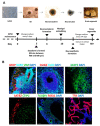
Figure 1
Figure 1
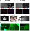
Generation of human brain organoids. (A) Protocol used for the generation of brain organoids. EB: embryoid body; hiPSC: human induced pluripotent stem cells. (B) Formation of brain structures was assessed by detection of different markers by immunofluorescence: Glial fibrillary acidic protein (GFAP), SOX2, Vimentin (VIM), OLIG2, Beta-III-tubulin (TUJ1), MAP2, SATB2, CTIP2, Tubulin-beta-IV (TUBβIV) and transthyretin (TTR). Scale bar: 100 µm.
Figure 2
Figure 2
Gz-NSC transplantation in brain organoids. (A) Gz-NSC were transduced with a lentiviral vector containing EGFP with a NLS to allow the detection of transplanted cells in the human tissue. Upper panels show phase contrast images and fluorescent images of EGFP-NLS. Lower panels show expression of the NSC markers Nestin and SOX2 and the ventral regional transcription factor NKX2.1 which is expressed at the level of the medial ganglionic eminence. The two Gz-NSC lines used for transplantation experiments expressed different levels of NKX2.1. Scale bar: 100 µm. (B) Coculture of Gz-NSC-EGFP and brain organoids. Gz-NSC EGFP form a neurosphere that fuses with the organoid. Scale bar: 100 µm. (C) Cell injection under a stereomicroscope inside a laminar flow cabinet. Scale bar: 100 µm and 1 mm in the inset (D) Detection of injected EGFP+ cells at different time points under an inverted fluorescence microscope. Scale bar: 100 µm and 50 µm in the insets.
Figure 3
Figure 3
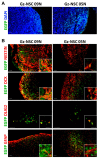
Gz-NSC survive and differentiate in the human brain organoids. (A) Survival and localization of two different lines of human Gz-NSC (EGFP+) transplanted into brain organoids. (B) Some transplanted cells maintain the stem cell phenotype as shown by the expression of Nestin while other cells differentiate into doublecortin (DCX)+ neuroblasts, OLIG2+ oligodendrocyte precursors and glial fibrillary acid (GFAP)+ astrocytes. Insets show colocalization of EGFP in green and the different markers in red. Scale bar: 50 µm and 20 µm in the insets.
Figure 4
Figure 4
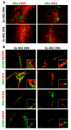
Gz-NSC survive and differentiate in the brain of immunodeficient mice. (A) Transplantation site of two different lines of human Gz-NSC identified by the expression of human nuclear antigen (HNA in green), surrounded by mouse reactive GFAP+ astrocytes and IBA1+ microglia (in red). (B) Some transplanted cells maintain a stem cell phenotype as shown by the expression of Nestin while other cells differentiate to DCX+ neuroblasts, OLIG2+ oligodendrocyte precursors and human specific (h)GFAP+ astrocytes. Insets show colocalization of nuclear human markers, HNA or KU80 respectively, in green, with the differentiation markers in red. Scale bar: 100 µm and 20 µm in the insets.
Figure 5
Figure 5
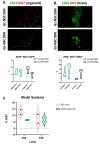
Gz-NSC proliferation rate after transplantation in the human brain organoids and in mice brain. (A) Gz-NSC proliferate within the brain organoids as shown by the expression of the proliferation marker KI67 in EGFP+ grafted cells 3 weeks after transplantation. Scale bar: 100 µm. (B) Gz-NSC proliferate at 3 weeks after transplantation in mice brain as shown by expression of the proliferation marker KI67 in HNA+ grafted cells. (C) Comparison of two Gz-NSC lines proliferation rate in human organoid versus mouse model.

References

    1. Bashor C.J., Hilton I.B., Bundukwala H., Smith D.M., Veiseh O. Engineering the next generation of cell-based therapeutics. Nat. Rev. Drug Discov. 2022;21:655–675. doi: 10.1038/s41573-022-00476-6. - DOI - PMC - PubMed
    1. Mak I.W.Y., Evaniew N., Ghert M. Lost in translation: Animal models and clinical trials in cancer treatment. Am. J. Transl. Res. 2014;6:114. - PMC - PubMed
    1. Ransohoff R.M. All (animal) models (of neurodegeneration) are wrong. Are they also useful? J. Exp. Med. 2018;215:2955–2958. doi: 10.1084/jem.20182042. - DOI - PMC - PubMed
    1. Donega V., Nijboer C.H., Van Velthoven C.T.J., Youssef S.A., De Bruin A., Van Bel F., Kavelaars A., Heijnen C.J. Assessment of long-term safety and efficacy of intranasal mesenchymal stem cell treatment for neonatal brain injury in the mouse. Pediatr. Res. 2015;78:520–526. doi: 10.1038/pr.2015.145. - DOI - PMC - PubMed
    1. Nabar N.R., Yuan F., Lin X., Wang L., Bai G., Mayl J., Li Y., Zhou S.F., Wang J., Cai J., et al. Cell Therapy: A Safe and Efficacious Therapeutic Treatment for Alzheimer’s Disease in APP+PS1 Mice. PLoS ONE. 2012;15:e0243343. doi: 10.1371/journal.pone.0049468. - DOI - PMC - PubMed