

Exosomes Derived from Yak Follicular Fluid Increase 2-Hydroxyestradiol Secretion by Activating Autophagy in Cumulus Cells
- PMID: 36428401
- PMCID: PMC9686841
- DOI: 10.3390/ani12223174
Exosomes Derived from Yak Follicular Fluid Increase 2-Hydroxyestradiol Secretion by Activating Autophagy in Cumulus Cells
Abstract
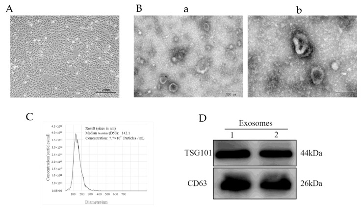
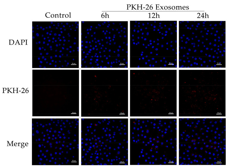
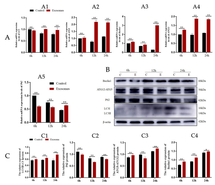
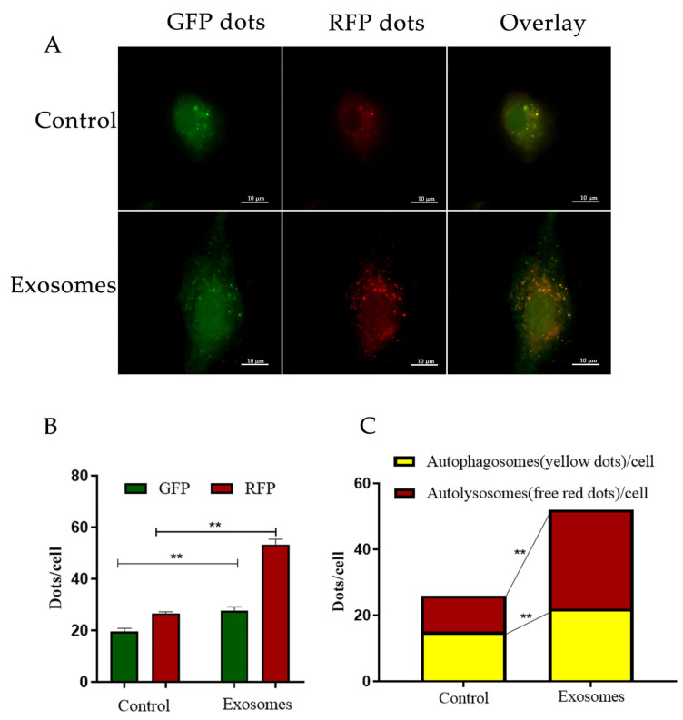
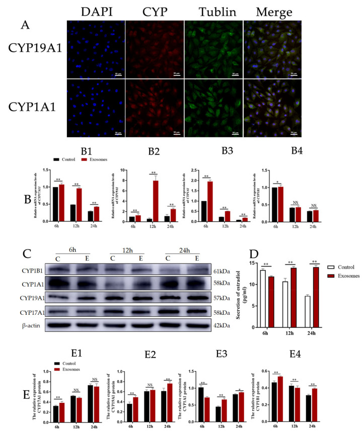
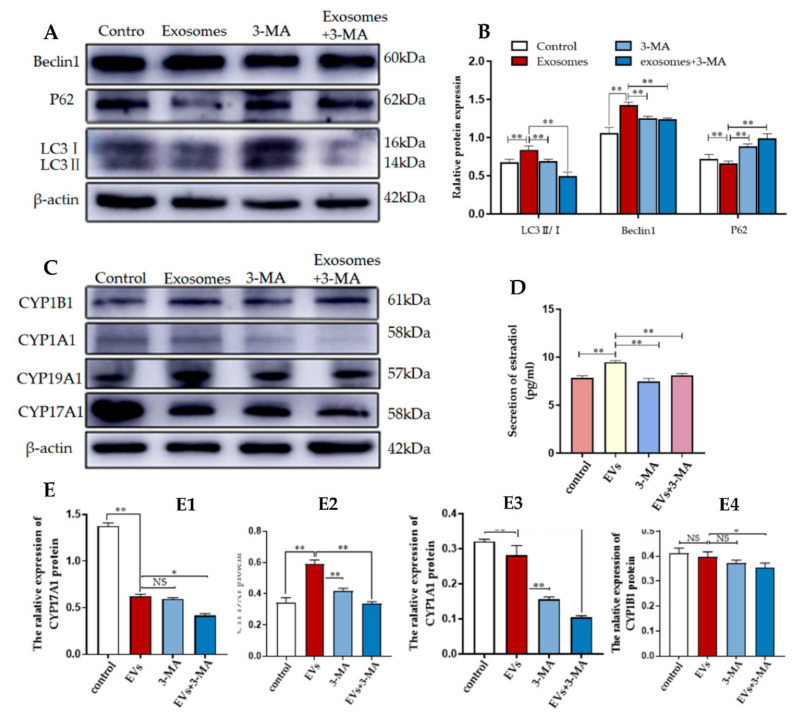
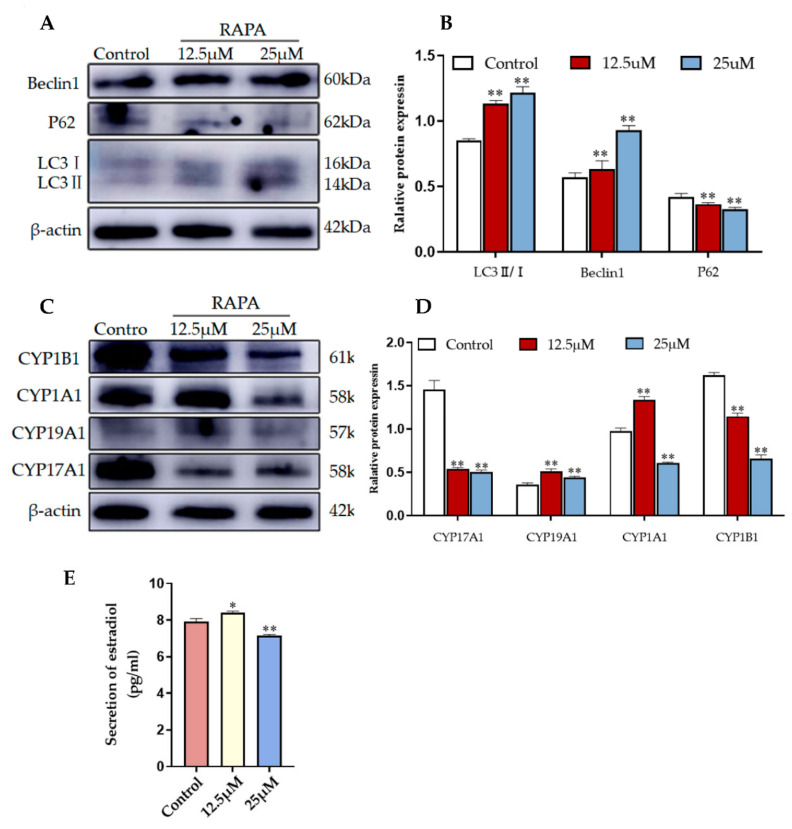
Exosomes in the follicular fluid can carry and transfer regulatory molecules to recipient cells, thus influencing their biological functions. However, the specific effects of yak follicular fluid exosomes on 2-hydroxyestrodiol (2-OHE2) secretion remain unknown. Here, we investigated whether yak follicular fluid exosomes can increase 2-OHE2 secretion through the activation of autophagy in cumulus cells (YCCs). In vitro cultured YCCs were treated with yak follicular fluid exosomes for 6, 12, and 24 h. The effects of yak follicular fluid exosomes on autophagy and 2-OHE2 secretion were evaluated through real-time quantitative fluorescence PCR (RT-qPCR), Western blotting (WB), transfected with RFP-GFP-LC3, immunohistochemistry, and ELISA. To further investigate whether 2-OHE2 secretion was related to autophagy, YCCs were administered with yak follicular fluid exosomes, 3-methyladenine (3-MA), and rapamycin (RAPA). The results revealed that treatment with yak follicular fluid exosomes activated autophagy in YCCs and increased 2-OHE2 secretion. Conversely, the inhibition of autophagy with 3-MA blocked these effects, suggesting that autophagy has an important role in 2-OHE2 secretion in YCCs. Treatment of YCCs with rapamycin showed similar results with yak follicular fluid exosomes as there was an increase in 2-OHE2 secretion due to the activation of autophagy in the treated cumulus cells. Our results demonstrate that autophagy is enhanced by yak follicular fluid exosomes, and this is associated with an increase in 2-OHE2 secretion in YCCs.
Keywords: 2-hydroxyestrondiol; autophagy; cumulus cells; yak follicular fluid exosomes.
Conflict of interest statement
The authors declare no conflict of interest.
Figures
Similar articles
-
Follicular fluid exosomes regulate OVGP1 secretion in yak oviduct epithelial cells via autophagy in vitro.J Cell Physiol. 2023 May;238(5):1020-1035. doi: 10.1002/jcp.30988. Epub 2023 Apr 4. J Cell Physiol. 2023. PMID: 37013674
-
Epidermal growth factor regulates autophagy activity and endocytosis of yak cumulus cells in a concentration-dependent manner.Front Vet Sci. 2023 Jan 9;9:1081643. doi: 10.3389/fvets.2022.1081643. eCollection 2022. Front Vet Sci. 2023. PMID: 36699336 Free PMC article.
-
Exosomal and Non-Exosomal Transport of Extra-Cellular microRNAs in Follicular Fluid: Implications for Bovine Oocyte Developmental Competence.PLoS One. 2013 Nov 4;8(11):e78505. doi: 10.1371/journal.pone.0078505. eCollection 2013. PLoS One. 2013. PMID: 24223816 Free PMC article.
-
Follicular fluid exosomes act on the bovine oocyte to improve oocyte competence to support development and survival to heat shock.Reprod Fertil Dev. 2019 Apr;31(5):888-897. doi: 10.1071/RD18450. Reprod Fertil Dev. 2019. PMID: 30760387
-
[Exosomes as a new approach into cell-to-cell communication within the mammalian ovary].Postepy Biochem. 2019 Oct 24;65(4):263-270. doi: 10.18388/pb.2019_276. Postepy Biochem. 2019. PMID: 31945280 Review. Polish.
Cited by
-
CIRBP Enhances the Function of Yak Cumulus Cells by Activating AMPK/mTOR-Mediated Mitophagy.Biomolecules. 2025 May 24;15(6):759. doi: 10.3390/biom15060759. Biomolecules. 2025. PMID: 40563401 Free PMC article.
-
Hypoxia-Inducible Factor 1α Affects Yak Oocyte Maturation and Early Embryonic Development by Regulating Autophagy.Antioxidants (Basel). 2024 Jul 14;13(7):840. doi: 10.3390/antiox13070840. Antioxidants (Basel). 2024. PMID: 39061908 Free PMC article.
References
-
- Gu X., Li Y., Chen K., Wang X., Wang Z., Lian H., Lin Y., Rong X., Chu M., Lin J., et al. Exosomes derived from umbilical cord mesenchymal stem cells alleviate viral myocarditis through activating AMPK/mTOR-mediated autophagy flux pathway. J. Cell. Mol. Med. 2020;24:7515–7530. doi: 10.1111/jcmm.15378. - DOI - PMC - PubMed
Grants and funding
- No. 32160850,32160859/National Natural Science Foundation of China
- 20JR10RA561/Distinguished Young Scholars of Gansu Province
- 2022-0623-RCC-0307/Key Talent Project of Gansu Province
- No.20200624RCC0022/Innovative Talent Project of Gansu Province
- (Grant No. 21ZD10NA001)/The Science and Technology Major Project of Gansu Province
LinkOut - more resources
Full Text Sources
