

Estimation of Early Postmortem Interval from Long Noncoding RNA Gene Expression in the Incised Cutaneous Wound: An Experimental Study
- PMID: 36428487
- PMCID: PMC9687757
- DOI: 10.3390/biomedicines10112919
Estimation of Early Postmortem Interval from Long Noncoding RNA Gene Expression in the Incised Cutaneous Wound: An Experimental Study
Abstract
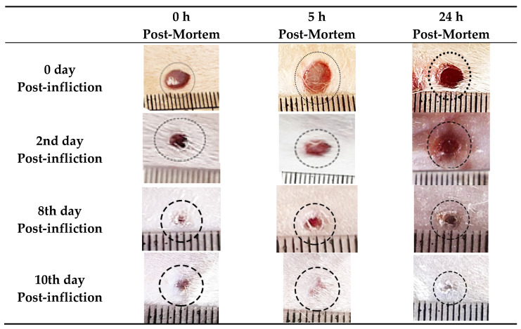
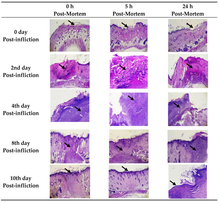
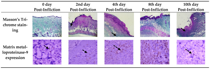
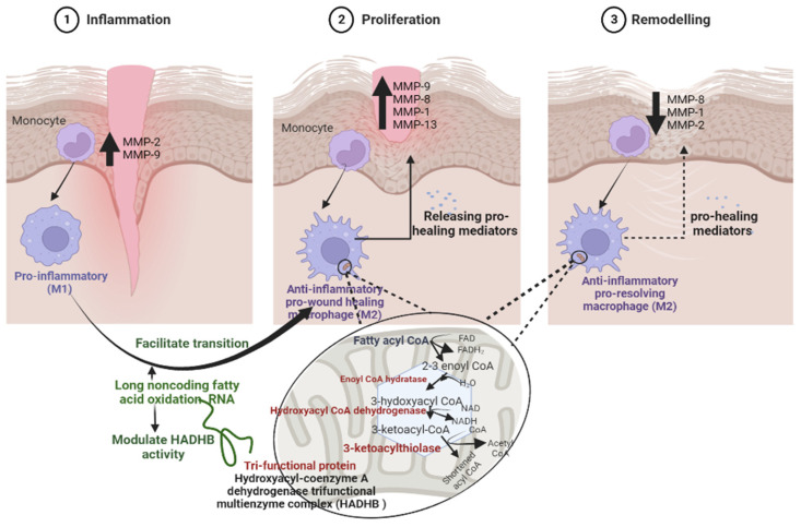
The assessment of alteration of postmortem RNA expression has forensic significance in estimating postmortem interval. To evaluate wound healing progression and the effect of different postmortem intervals, histopathological changes, immunohistochemical matrix metalloproteinase-9 (MMP-9) expression, and long noncoding fatty acid oxidation (lncFAO), RNA expression was assessed in the incised cutaneous wound model. A full-thickness cutaneous wound was inflicted on 75 rats. All 15 rats were sacrificed at different post-infliction intervals (0, 2, 4, 8 and 10 days), and the cutaneous wounds (n = 5) were excised at different postmortem intervals (0, 5, and 24 h after euthanasia). The maximal inflammatory healing stage was detected at day 4 post-infliction, while near complete healing, thick mature collagen deposition was detected at day 10 post-infliction. LncFAO expression was significantly over-expressed with increasing wound age. MMP-9 was detectable on injury day with continuous elevation until 8 days post-wounding, which later decreased. Although histopathological and immunohistochemical examinations within 24 h postmortem did not show any remarkable changes, lncFAO RNA expression showed a significant negative correlation with hours passed since death. The combined use of histopathological changes, immunohistochemical expression of MMP-9, and molecular expression of lncFAO could be appropriate in wound dating verification. Among these factors, lncFAO could be a reliable indicator in postmortem interval estimation.
Keywords: forensic medicine; incised wound; long non-coding RNA; matrix metalloproteinase-9; postmortem interval.
Conflict of interest statement
The authors declare no conflict of interest.
Figures

Similar articles
-
Expression of matrix metalloproteinase-9 (MMP-9) in human skin within 1 hour after injury through immunohistochemical staining: a pilot study.Int J Legal Med. 2024 Sep;138(5):1985-1990. doi: 10.1007/s00414-024-03243-x. Epub 2024 May 1. Int J Legal Med. 2024. PMID: 38691159
-
Complex challenges of estimating the age and vitality of muscle wounds: a study with matrix metalloproteinases and their inhibitors on animal and human tissue samples.Int J Legal Med. 2021 Sep;135(5):1843-1853. doi: 10.1007/s00414-021-02563-6. Epub 2021 May 26. Int J Legal Med. 2021. PMID: 34041592 Free PMC article.
-
Topical application of Acalypha indica accelerates rat cutaneous wound healing by up-regulating the expression of Type I and III collagen.J Ethnopharmacol. 2012 Jun 26;142(1):14-22. doi: 10.1016/j.jep.2012.04.005. Epub 2012 Apr 14. J Ethnopharmacol. 2012. PMID: 22521732
-
Propranolol improves cutaneous wound healing in streptozotocin-induced diabetic rats.Eur J Pharmacol. 2009 Jun 2;611(1-3):77-84. doi: 10.1016/j.ejphar.2009.03.053. Epub 2009 Apr 1. Eur J Pharmacol. 2009. PMID: 19344703
-
Immunohistochemical parameters for the age estimation of human skin wounds. A review.Am J Forensic Med Pathol. 1995 Sep;16(3):203-9. doi: 10.1097/00000433-199509000-00003. Am J Forensic Med Pathol. 1995. PMID: 7495259 Review.
Cited by
-
Are pre-analytical factors fully considered in forensic FFPE molecular analyses? A systematic review reveals the need for standardised procedures.Int J Legal Med. 2025 Jul;139(4):1439-1452. doi: 10.1007/s00414-025-03480-8. Epub 2025 Apr 2. Int J Legal Med. 2025. PMID: 40172636 Free PMC article.
-
Decoding Time of Death: Histopathological Dynamics of Intervertebral Discs as a Novel Marker for Postmortem Interval Estimation.Diagnostics (Basel). 2025 Mar 2;15(5):605. doi: 10.3390/diagnostics15050605. Diagnostics (Basel). 2025. PMID: 40075852 Free PMC article.
-
An experimental study to estimate the early postmortem interval based on the degradation of lncRNAs in rat brain tissue.Sci Rep. 2024 Aug 23;14(1):19586. doi: 10.1038/s41598-024-70678-y. Sci Rep. 2024. PMID: 39179611 Free PMC article.
References
-
- Nakayama Y., Fujiu K., Yuki R., Oishi Y., Morioka M.S., Isagawa T., Matsuda J., Oshima T., Matsubara T., Sugita J., et al. A long noncoding RNA regulates inflammation resolution by mouse macrophages through fatty acid oxidation activation. Proc. Natl. Acad. Sci. USA. 2020;117:14365–14375. doi: 10.1073/pnas.2005924117. - DOI - PMC - PubMed
LinkOut - more resources
Full Text Sources
Miscellaneous
