Review
. 2022 Nov 21;10(11):2988.
doi: 10.3390/biomedicines10112988.

Cancer Metastasis and Treatment Resistance: Mechanistic Insights and Therapeutic Targeting of Cancer Stem Cells and the Tumor Microenvironment

Affiliations
Review

Cancer Metastasis and Treatment Resistance: Mechanistic Insights and Therapeutic Targeting of Cancer Stem Cells and the Tumor Microenvironment

Ethan J Kilmister et al. Biomedicines. .

Abstract

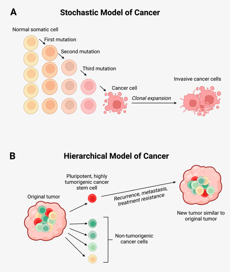
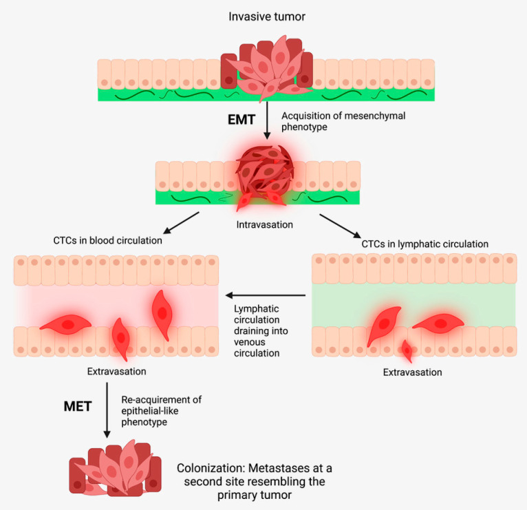
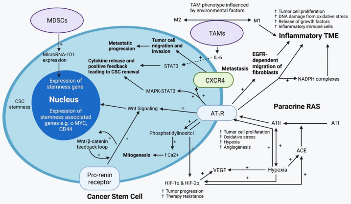
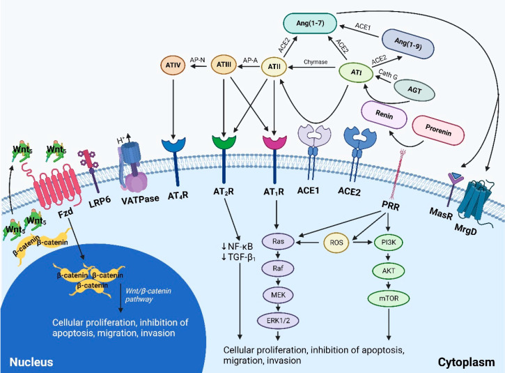
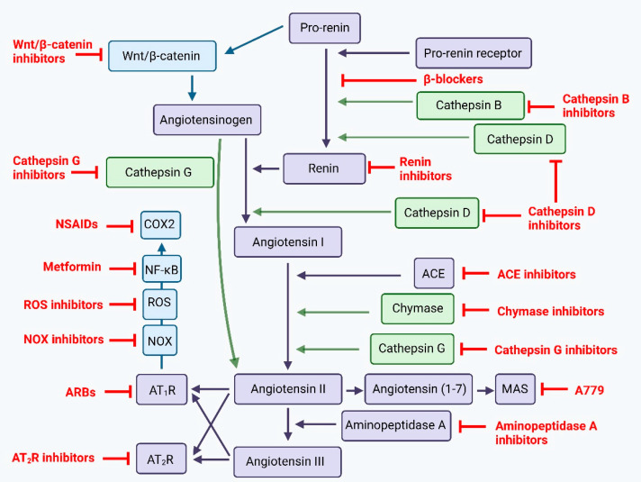
Cancer metastasis and treatment resistance are the main causes of treatment failure and cancer-related deaths. Their underlying mechanisms remain to be fully elucidated and have been attributed to the presence of cancer stem cells (CSCs)-a small population of highly tumorigenic cancer cells with pluripotency and self-renewal properties, at the apex of a cellular hierarchy. CSCs drive metastasis and treatment resistance and are sustained by a dynamic tumor microenvironment (TME). Numerous pathways mediate communication between CSCs and/or the surrounding TME. These include a paracrine renin-angiotensin system and its convergent signaling pathways, the immune system, and other signaling pathways including the Notch, Wnt/β-catenin, and Sonic Hedgehog pathways. Appreciation of the mechanisms underlying metastasis and treatment resistance, and the pathways that regulate CSCs and the TME, is essential for developing a durable treatment for cancer. Pre-clinical and clinical studies exploring single-point modulation of the pathways regulating CSCs and the surrounding TME, have yielded partial and sometimes negative results. This may be explained by the presence of uninhibited alternative signaling pathways. An effective treatment of cancer may require a multi-target strategy with multi-step inhibition of signaling pathways that regulate CSCs and the TME, in lieu of the long-standing pursuit of a 'silver-bullet' single-target approach.

Keywords: cancer stem cell; metastasis; renin-angiotensin system; treatment resistance; tumor microenvironment.


Conflict of interest statement

The authors declare no conflict of interest.

Figures

Figure 1
Figure 1
(A) The stochastic model of cancer proposes that a normal somatic cell accumulates oncogenic mutations in a stepwise manner and becomes a cancer cell that undergoes clonal expansion to form a tumor. (B) The hierarchical model of cancer proposes the presence of a highly tumorigenic cancer stem cell (CSC) sitting atop the tumor cellular hierarchy and divides asymmetrically to form non-tumorigenic cancer cells that form the bulk of the tumor, and identical CSCs that form new tumors like the original tumor. Adapted from the Atlas of Extreme Facial Cancer, Springer Nature [9]. Diagram recreated with BioRender.com, accessed on 1 November 2022.
Figure 2
Figure 2
In cancer metastasis, tumor cells undergo epithelial-mesenchymal transition (EMT) to form mesenchymal-like cells, which undergo intravasation to enter the blood and/or lymphatic circulations as circulating tumor cells (CTCs). CTCs then undergo mesenchymal-epithelial transition (MET) and extravasate into distant tissue sites, where metastases may form. Adapted from the Atlas of Extreme Facial Cancer, Springer Nature, 2022 [9]. Diagram recreated with BioRender.com, accessed on 1 November 2022.
Figure 3
Figure 3
A cancer stem cell (CSC) residing within the tumor microenvironment (TME) which is regulated by the paracrine renin–angiotensin system (RAS) and the immune system. The active end-product of the paracrine RAS—angiotensin II (ATII), activates ATII receptor 1 (AT1R) resulting in increased tumor cell proliferation, oxidative stress, hypoxia and angiogenesis, and inflammation—the hallmarks of cancer. This contributes to an inflammatory TME by increasing the number of inflammatory cells, partly by increasing the number of NADPH complexes, leading to tumor cell proliferation, DNA damage from oxidative stress, and release of growth factors. AT1R also activates phosphatidylinositol signaling which increases cytosolic Ca2+ to promote mitogenesis. Hypoxia increases paracrine RAS activity by up-regulating angiotensin-converting enzyme (ACE) and hypoxia-inducible factor 1α (HIF-1α) and HIF-2α expression, which increase tumor progression and treatment resistance. HIF-1α, HIF-2α, and hypoxia also increase the expression of vascular endothelial growth factor (VEGF) which promotes angiogenesis. Binding of AT1R to C-X-C chemokine receptor type 4 (CXCR4) promotes tumor cell migration and invasion, leading to metastatic spread. AT1R, via MAPK-STAT3 signaling, contributes to a cytokine release that leads to CSC renewal. AT1R signaling also contributes to the migration of fibroblasts in an epidermal growth factor receptor (EGFR)-dependent fashion. AT1R signaling and the pro-renin receptor, acting in a feedback loop with Wnt/β-catenin, increase Wnt signaling which promotes CSC stemness by up-regulating stemness-associated markers. Myeloid-derived suppressor cells (MDSCs) promote CSC characteristics by increasing the expression of microRNA-101 that induces expression of stemness-related genes in CSCs. Under the influence of the TME, polarization of tumor-associated macrophages (TAMs) within the TME, changes from the M1 to M2 phenotype. M2 TAMs induce the proliferation of CSCs via interleukin-6 (IL-6)-induced activation of STAT3, leading to cytokine release and positive feedback that contribute to CSC renewal. Abbreviations: ATI, angiotensin I; AT2R, ATII receptor 2; Ang1–7, angiotensin 1–7; ATIII, angiotensin III; MAPK, mitogen-activated protein kinase. Adapted from the Journal of Histochemistry and Cytochemistry [90]. Diagram recreated with BioRender.com, accessed on 1 November 2022.
Figure 4
Figure 4
A schema showing the effect of the paracrine renin–angiotensin system (RAS) and its convergent signaling pathways on the tumor microenvironment to influence cellular proliferation, invasiveness, and cell survival in cancer development. The RAS interacts with downstream pathways, such as the Ras/RAF/MEK/ERK (light blue) pathway and the PI3K/AKT/mTOR (dark blue) pathway, and the up-stream Wnt/β-catenin pathway (intermediate blue) that influence cellular proliferation, migration, inhibition of apoptosis, migration, and invasion (see text). PRR, pro-renin receptor; LRP6, low-density lipoprotein receptor-related protein; Fzd, frizzled receptor; Cath G, cathepsin G; Cath B, cathepsin B; Cath D, cathepsin D; ACE1, angiotensin-converting enzyme 1; ACE2, angiotensin-converting enzyme 2; ADP, adenosine diphosphate; AGT, angiotensinogen; ATP, adenosine triphosphate; Ang(1–7), angiotensin (1–7); Ang(1–9), angiotensin (1–9); AP-A, aminopeptidase-A; NEP, neutral endopeptidase; AP-N, aminopeptidase-N; ATI, angiotensin I; ATII, angiotensin II; ATIII, angiotensin III; ATIV, angiotensin IV; AT1R, angiotensin II receptor 1; AT2R, angiotensin II receptor 2; AT4R, angiotensin II receptor 4; MrgD, Mas-related-G protein coupled receptor; MasR, Mas receptor; mTOR, mammalian target of rapamycin; NF-κB, nuclear factor kappa B; TGF-β1, transforming growth factor-β1; V-ATPase, vacuolar H+-adenosine triphosphate. Adapted from Cancers [134]. Diagram recreated with BioRender.com, accessed on 1 November 2022.
Figure 5
Figure 5
The renin-angiotensin system and its bypass loops and converging signaling pathways can be targeted at different points. The renin-angiotensin system (black) regulates blood pressure, stem cell differentiation, and tumor development. Bypass loops in the system involving cathepsins and chymase (green) provide redundancy, while convergent inflammatory and development signaling pathways (blue) have multiple roles and effects. Multiple points of the pathway can be targeted by specific inhibitors (red). ACE, angiotensin converting enzyme; ARBs, AT1R blockers; ROS, reactive oxygen species; NSAIDS, non-steroidal anti-inflammatory drugs. Adapted from Frontiers in Oncology [120]. Diagram recreated with BioRender.com, accessed on 1 November 2022.

References

    1. Lambert A.W., Pattabiraman D.R., Weinberg R.A. Emerging biological principles of metastasis. Cell. 2017;168:670–691. doi: 10.1016/j.cell.2016.11.037. - DOI - PMC - PubMed
    1. Dillekås H., Rogers M.S., Straume O. Are 90% of deaths from cancer caused by metastases? Cancer Med. 2019;8:5574–5576. doi: 10.1002/cam4.2474. - DOI - PMC - PubMed
    1. Steeg P.S. Targeting metastasis. Nat. Rev. Cancer. 2016;16:201–218. doi: 10.1038/nrc.2016.25. - DOI - PMC - PubMed
    1. Chaffer C.L., Weinberg R.A. A perspective on cancer cell metastasis. Science. 2011;331:1559–1564. doi: 10.1126/science.1203543. - DOI - PubMed
    1. Fares J., Fares M.Y., Khachfe H.H., Salhab H.A., Fares Y. Molecular principles of metastasis: A hallmark of cancer revisited. Signal Transduct Target Ther. 2020;5:28. doi: 10.1038/s41392-020-0134-x. - DOI - PMC - PubMed

LinkOut - more resources