Profiling the Adrenergic System in Breast Cancer and the Development of Metastasis
- PMID: 36428611
- PMCID: PMC9688855
- DOI: 10.3390/cancers14225518
Profiling the Adrenergic System in Breast Cancer and the Development of Metastasis
Abstract
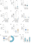
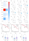
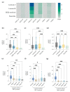
Epidemiological studies and preclinical models suggest that chronic stress might accelerate breast cancer (BC) growth and the development of metastasis via sympathetic neural mechanisms. Nevertheless, the role of each adrenergic pathway (α1, α2, and β) in human samples remains poorly depicted. Herein, we propose to characterize the profile of the sympathetic system (e.g., release of catecholamines, expression of catecholamine metabolic enzymes and adrenoreceptors) in BC patients, and ascertain its relevance in the development of distant metastasis. Our results demonstrated that BC patients exhibited increased plasma levels of catecholamines when compared with healthy donors, and this increase was more evident in BC patients with distant metastasis. Our analysis using the BC-TCGA database revealed that the genes coding the most expressed adrenoreceptors in breast tissues (ADRA2A, ADRA2C, and ADRB2, by order of expression) as well as the catecholamine synthesizing (PNMT) and degrading enzyme (MAO-A and MAO-B) genes were downregulated in BC tissues. Importantly, the expression of ADRA2A, ADRA2C, and ADRB2 was correlated with metastatic BC and BC subtypes, and thus the prognosis of the disease. Overall, we gathered evidence that under stressful conditions, both the α2- and β2-signaling pathways might work on a synergetic matter, thus paving the way for the development of new therapeutic approaches.
Keywords: adrenoreceptors; bone metastasis; breast cancer; stress; sympathetic nervous system.
Conflict of interest statement
The authors declare no conflict of interest.
Figures


Similar articles
-
The Secretome of Parental and Bone Metastatic Breast Cancer Elicits Distinct Effects in Human Osteoclast Activity after Activation of β2 Adrenergic Signaling.Biomolecules. 2023 Mar 30;13(4):622. doi: 10.3390/biom13040622. Biomolecules. 2023. PMID: 37189370 Free PMC article.
-
Prognostic significance of α- and β2-adrenoceptor gene expression in breast cancer patients.Br J Clin Pharmacol. 2019 Sep;85(9):2143-2154. doi: 10.1111/bcp.14030. Epub 2019 Jul 31. Br J Clin Pharmacol. 2019. PMID: 31218733 Free PMC article.
-
α2-Adrenergic blockade mimics the enhancing effect of chronic stress on breast cancer progression.Psychoneuroendocrinology. 2015 Jan;51:262-70. doi: 10.1016/j.psyneuen.2014.10.004. Epub 2014 Oct 12. Psychoneuroendocrinology. 2015. PMID: 25462899 Free PMC article.
-
Adrenergic receptors and cardiovascular effects of catecholamines.Ann Endocrinol (Paris). 2021 Jun;82(3-4):193-197. doi: 10.1016/j.ando.2020.03.012. Epub 2020 Mar 18. Ann Endocrinol (Paris). 2021. PMID: 32473788 Review.
-
Stress in Metastatic Breast Cancer: To the Bone and Beyond.Cancers (Basel). 2022 Apr 8;14(8):1881. doi: 10.3390/cancers14081881. Cancers (Basel). 2022. PMID: 35454788 Free PMC article. Review.
Cited by
-
The Secretome of Parental and Bone Metastatic Breast Cancer Elicits Distinct Effects in Human Osteoclast Activity after Activation of β2 Adrenergic Signaling.Biomolecules. 2023 Mar 30;13(4):622. doi: 10.3390/biom13040622. Biomolecules. 2023. PMID: 37189370 Free PMC article.
-
The Expression of Trace Amine-Associated Receptors (TAARs) in Breast Cancer Is Coincident with the Expression of Neuroactive Ligand-Receptor Systems and Depends on Tumor Intrinsic Subtype.Biomolecules. 2023 Sep 7;13(9):1361. doi: 10.3390/biom13091361. Biomolecules. 2023. PMID: 37759760 Free PMC article.
-
Wrecking neutrophil extracellular traps and antagonizing cancer-associated neurotransmitters by interpenetrating network hydrogels prevent postsurgical cancer relapse and metastases.Bioact Mater. 2024 May 14;39:14-24. doi: 10.1016/j.bioactmat.2024.05.022. eCollection 2024 Sep. Bioact Mater. 2024. PMID: 38783926 Free PMC article.
-
Pan-cancer analysis of the role of α2C-adrenergic receptor (ADRA2C) in human tumors and validation in glioblastoma multiforme models.J Cancer. 2024 Sep 9;15(17):5691-5709. doi: 10.7150/jca.98240. eCollection 2024. J Cancer. 2024. PMID: 39308687 Free PMC article.
-
ADRA2A promotes the classical/progenitor subtype and reduces disease aggressiveness of pancreatic cancer.bioRxiv [Preprint]. 2024 Mar 13:2024.03.12.584316. doi: 10.1101/2024.03.12.584316. bioRxiv. 2024. Update in: Carcinogenesis. 2024 Nov 22;45(11):845-856. doi: 10.1093/carcin/bgae056. PMID: 38903083 Free PMC article. Updated. Preprint.
References
-
- Ferlay J.E.M., Lam F., Colombet M., Mery L., Piñeros M., Znaor A., Soerjomataram I., Bray F. Global Cancer Observatory: Cancer Today. Lyon, France: International Agency for Research on Cancer. 2020. [(accessed on 12 January 2021)]. Available online: https://gco.iarc.fr/today.
-
- Zielonke N., Gini A., Jansen E.E.L., Anttila A., Segnan N., Ponti A., Veerus P., de Koning H.J., van Ravesteyn N.T., Heijnsdijk E.A.M. Evidence for reducing cancer-specific mortality due to screening for breast cancer in Europe: A systematic review. Eur. J. Cancer Oxf. Engl. 1990. 2020;127:191–206. doi: 10.1016/j.ejca.2019.12.010. - DOI - PubMed
Grants and funding
LinkOut - more resources
Full Text Sources
Research Materials
Miscellaneous

