. 2022 Nov 17;14(22):5648.
doi: 10.3390/cancers14225648.

Regression of Lung Cancer in Mice by Intranasal Administration of SARS-CoV-2 Spike S1

Affiliations

Regression of Lung Cancer in Mice by Intranasal Administration of SARS-CoV-2 Spike S1

Monica Sheinin et al. Cancers (Basel). .

Abstract

This study underlines the importance of SARS-CoV-2 spike S1 in prompting death in cultured non-small cell lung cancer (NSCLC) cells and in vivo in lung tumors in mice. Interestingly, we found that recombinant spike S1 treatment at very low doses led to death of human A549 NSCLC cells. On the other hand, boiled recombinant SARS-CoV-2 spike S1 remained unable to induce death, suggesting that the induction of cell death in A549 cells was due to native SARS-CoV-2 spike S1 protein. SARS-CoV-2 spike S1-induced A549 cell death was also inhibited by neutralizing antibodies against spike S1 and ACE2. Moreover, our newly designed wild type ACE2-interacting domain of SARS-CoV-2 (wtAIDS), but not mAIDS, peptide also attenuated SARS-CoV-2 spike S1-induced cell death, suggesting that SARS-CoV-2 spike S1-induced death in A549 NSCLC cells depends on its interaction with ACE2 receptor. Similarly, recombinant spike S1 treatment also led to death of human H1299 and H358 NSCLC cells. Finally, 4-(methylnitrosamino)-1-(3-pyridyl)-1-butanone (NNK) intoxication led to the formation tumors in lungs of A/J mice and alternate day intranasal treatment with low dose of recombinant SARS-CoV-2 spike S1 from 22-weeks of NNK insult (late stage) induced apoptosis and tumor regression in the lungs. These studies indicate that SARS-CoV-2 spike S1 may have implications for lung cancer treatment.

Keywords: ACE2; NNK mouse model of lung cancer; SARS-CoV-2 spike S1; apoptosis; human lung cancer cells.


Conflict of interest statement

The authors declare no conflict of interest.

Figures

Figure 1
Figure 1
Time line for LDH and MTT assays.
Figure 2
Figure 2
Effect of recombinant SARS-CoV-2 spike S1 on the survival of human A549 lung cancer cells. A549 cells were treated with different concentrations (1, 5, and 10 ng/mL) of recombinant spike S1 protein for 24 h under serum-free condition followed by monitoring cell death by LDH release (A) and MTT (B). FACS double staining with annexin V and propidium iodide (PI) was also performed (C). Quantitative analysis of percent apoptotic cells is presented (D). Values are presented as mean ± SD of three independent experiments. * p < 0.05; ** p < 0.01.
Figure 3
Figure 3
Recombinant SARS-CoV-2 spike S1 induces apoptosis in human A549 lung cancer cells. A549 cells were treated with different doses of spike S1 protein for 12 h under serum-free condition followed by monitoring apoptosis by TUNEL (A). TUNEL positive cells were counted in 10 varied images per group and plotted as percent of total cells (B). (C) Cells were immunoblotted for apoptosis-related molecules (BAD, caspase 3 and cleaved caspase 3). Actin was run as a loading control. For original blots, please see Figure S1. Bands were scanned and values ((D), BAD/Actin; (E) cleaved caspase 3/Actin; (F) caspase 3/Actin) presented as relative to control. (G) Cells were immunoblotted for survival-related molecule (Bcl2). Actin was run as a loading control. (H) Bands were scanned and values (Bcl2/Actin) presented as relative to control. Results are mean ± SD of three different experiments. * p < 0.05; ** p < 0.01; *** p < 0.001; ns, not significant. Scale bar = 22 µm.
Figure 4
Figure 4
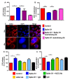
Spike S1-mediated death of human A549 lung cancer cells depends on ACE2 receptor. A549 cells were treated with 5 ng/mL spike S1 protein in the presence or absence of neutralizing antibodies against spike S1 (0.5 µg/mL) under serum-free condition. After 24 h, cell viability was monitored by LDH release (A) and MTT (B). Control A549 cells were immunostained for ACE2 (C). DAPI was used to stain nuclei. Cells were treated with 5 ng/mL spike S1 protein in the presence or absence of neutralizing antibodies against ACE2 (0.5 µg/mL) under serum-free condition. After 24 h, cell viability was monitored by LDH release (D) and MTT (E). Results are mean ± SD of three different experiments. * p < 0.05; ** p < 0.01; *** p< 0.001. Scale bar = 10 µm.
Figure 5
Figure 5
Selective disruption of ACE2-to-spike S1 interaction reduces spike S1-induced death in human A549 lung cancer cells. A549 cells were treated with 5 ng/mL spike S1 protein in the presence or absence of different concentrations of wtAIDS and mAIDS peptides under serum-free condition. After 24 h, cell viability was monitored by LDH release (A) and MTT (B). After 12 h of treatment, apoptosis was monitored by TUNEL (C). TUNEL positive cells were counted in 10 varied images per group and plotted as percent of total cells (D). Results are mean ± SD of three different experiments. * p < 0.05; ** p < 0.01; *** p< 0.001. Scale bar = 22 µm.
Figure 6
Figure 6
Effect of recombinant SARS-CoV-2 spike S1 on the survival of human H1299 and H358 lung cancer cells. H1299 (A,C) and H358 (B,D) cells were treated with spike S1 protein for 24 h under serum-free condition followed by monitoring cell death by LDH release (A,B) and MTT (C,D). Results are mean + SD of three different experiments. * p < 0.05; ** p < 0.01; *** p< 0.001.
Figure 7
Figure 7
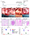
Intranasal administration of recombinant SARS-CoV-2 spike S1 causes regression of lung tumor in NNK-insulted female A/J mice. The experimental design is illustrated for NNK-induced lung cancer in A/J mice (A). Briefly, female A/J mice (5–6 week old) received two intraperitoneal (i.p.) injections of NNK (50 mg/kg body weight) one week apart. Tumor development was analyzed after 26 weeks of NNK intoxication. Mice were treated with spike S1 (50 ng/mouse/2 d) intranasally on alternate days starting from 22 weeks of NNK insult for 4 weeks followed by sacrificing mice on 26 weeks. Representative lung appearance in different groups of mice (B). Lung sections were stained for H&E (C). The histological tumor area was quantified in a 4x field as a percent of control (D). The number of lung lesions is shown in different groups of mice (E). Results are mean ± SD of 5 mice per group. *** p< 0.001; NS, not significant. Scale bar = 20 µm.
Figure 8
Figure 8
Intranasal administration of recombinant SARS-CoV-2 spike S1 induces apoptosis in lung tumors of NNK-insulted female A/J mice. Female A/J mice (5–6 week old) received two intraperitoneal (i.p.) injections of NNK (50 mg/kg body weight) one week apart. Mice were treated with spike S1 (50 ng/mouse/2 d) intranasally on alternate days starting from 22 weeks of NNK insult for 4 weeks followed by sacrificing mice on 26 weeks. Tumor tissue sections were labeled for TUNEL (A) followed by counting of TUNEL-positive cells in two sections (two images per section) of each of five mice per group (B). Results are mean + SD of 5 mice per group. *** p < 0.001; NS, not significant. Scale bar = 40 µm.

References

    1. Centers for Disease Control Chronic disease reports: Deaths from lung cancer--United States, 1986. JAMA. 1989;262:1170–1172. - PubMed
    1. Dela Cruz C.S., Tanoue L.T., Matthay R.A. Lung Cancer: Epidemiology, Etiology, and Prevention. Clin. Chest Med. 2011;32:605–644. doi: 10.1016/j.ccm.2011.09.001. - DOI - PMC - PubMed
    1. Siegel R.L., Miller K.D., Fuchs H.E., Jemal A. Cancer statistics, 2022. CA A Cancer J. Clin. 2022;72:7–33. doi: 10.3322/caac.21708. - DOI - PubMed
    1. Rachow T., Schiffl H., Lang S.M. Risk of lung cancer and renin–angiotensin blockade: A concise review. J. Cancer Res. Clin. Oncol. 2020;147:195–204. doi: 10.1007/s00432-020-03445-x. - DOI - PMC - PubMed
    1. Alexander M., Kim S.Y., Cheng H. Update 2020: Management of Non-Small Cell Lung Cancer. Lung. 2020;198:897–907. doi: 10.1007/s00408-020-00407-5. - DOI - PMC - PubMed

LinkOut - more resources