. 2022 Nov 21;14(22):5710.
doi: 10.3390/cancers14225710.

Tat-hspb1 Suppresses Clear Cell Renal Cell Carcinoma (ccRCC) Growth via Lysosomal Membrane Permeabilization

Affiliations

Tat-hspb1 Suppresses Clear Cell Renal Cell Carcinoma (ccRCC) Growth via Lysosomal Membrane Permeabilization

Lin Zhang et al. Cancers (Basel). .

Abstract

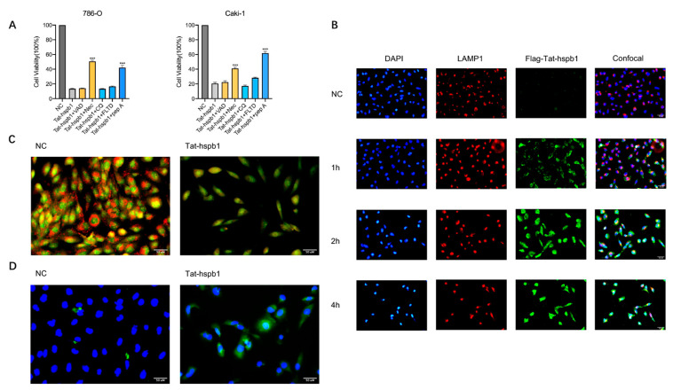
Clear cell renal cell carcinoma (ccRCC) is the most prevalent kidney cancer, of which the incidence is increasing worldwide with a high mortality rate. Bioactive peptides are considered a significant class of natural medicines. We applied mass spectrometry-based peptidomic analysis to explore the peptide profile of human renal clear cell carcinoma and adjacent normal tissues. A total of 18,031 peptides were identified, of which 105 unique peptides were differentially expressed (44 were up-regulated and 61 were down-regulated in ccRCC tissues). Through bioinformatic analysis, we finally selected one peptide derived from the HSPB1 protein (amino acids 12-35 of the N-terminal region of HSPB1). Next, we fused this peptide to the HIV-Tat, generated a novel peptide named Tat-hspb1, and found that Tat-hspb1 inhibited ccRCC cells' viability while being less cytotoxic to normal epithelial cells. Furthermore, Tat-hspb1 induced apoptosis and inhibited the proliferation and migration of ccRCC cells. Furthermore, we demonstrated that Tat-hspb1 was predominantly localized in lysosomes after entering the ccRCC cell and induced lysosomal membrane permeabilization (LMP) and the release of cathepsin D from lysosomes. Taken together, Tat-hspb1 has the potential to serve as a new anticancer drug candidate.

Keywords: apoptosis; lysosomal membrane permeabilization (LMP); peptide; renal cancer.


Conflict of interest statement

The authors declare no conflict of interest.

Figures

Figure 1
Figure 1
Characteristics of differentially expressed peptides and bioinformatic analysis of precursor proteins from which differentially expressed peptides were derived. (A) A volcano plot identified 115 differentially expressed peptides (fold change >2 or <0.5, p < 0.05). (B) A heat map identified 105 unique differentially expressed peptides. (C) Molecular function. (D) Biological processes. (E) Cellular component. (F) Reactome pathway enrichment analysis. (G) Protein–protein interaction network analysis based on STRING.
Figure 2
Figure 2
Selection of peptides with potential bioactivity. Cell viability was determined by CCK-8 assays. (A) A Venn diagram identified two peptides derived from the same precursor protein HSPB1. (BD) Renal cancer cell lines 786-O, Caki-1, and A498 and (E,F) normal epithelial cells HUVEC and HKC were treated with various concentrations of Tat-hspb1 for 24 h and 48 h or double-distilled water as a control. The results are expressed as the means ± SD of three independent experiments.
Figure 3
Figure 3
Tat-hspb1 inhibits proliferation and migration of renal cancer cells. (A) Representative images and quantification results of colony formation assay of 786-O and Caki-1 cells after treatment with low-gradient concentrations of Tat-hspb1. (B) Representative images and quantification results of wound-healing assay of 786-O and Caki-1 cells after treatment with low-gradient Tat-hspb1 concentrations. The results are expressed as the means ± SD of three independent experiments. *** p < 0.001.
Figure 4
Figure 4
Tat-hspb1 induces apoptosis in renal cancer cells. (A) Bright-field photomicrographs show obvious morphological change of 786-O and Caki-1 cells after treatment with Tat-hspb1 (80 μg/mL) or double-distilled water as a control. (B) 786-O and Caki-1 cells were treated with the different concentrations of Tat-hspb1 for 24 h, Representative graphs were obtained from cytometry analysis. (C) 786-O and Caki-1 cells were treated with the different concentrations of Tat-hspb1 for 24 h; the protein expression of caspase-3, cleaved-caspase-3, caspase-8, cleaved-caspase-8, caspase-9, and cleaved-caspase-9 was determined by Western Blot. The results are expressed as the means ± SD of three independent experiments. ** p < 0.01, *** p < 0.001.
Figure 5
Figure 5
Tat-hspb1 induces lysosomal membrane permeabilization (LMP) in renal cancer cells. (A) 786-O and Caki-1 cells were treated with Tat-hspb1 (80 μg/mL) either alone or combined with specific inhibitors, Z-VAD-FMK (80 μM), necrostatin-1 (80 μM), CQ (80 μM), Ac-FLTD-CMK (80 μM), and pepstatin-1 (80 μM) for 24 h, and viability was assessed by CCK-8 assay. (B) 786-O cells were treated with Flag-Tat-hspb1 (80 μg/mL) for 1, 2, and 4 h, and double-distilled water was used as a control. Typical confocal images were obtained, where green fluorescence represents Tat-hspb1 and red fluorescence represents LAMP1. (C) 786-O cells untreated or treated with Tat-hspb1 (80 μg/mL for 2 h) were stained with acridine orange (AO). (D) 786-O cells untreated or treated with Tat-hspb1 (80 μg/mL for 2 h) were stained with the anti-Cathepsin D antibody. The results are expressed as the means ± SD of three independent experiments. *** p < 0.001.

References

    1. Sung H., Ferlay J., Siegel R.L., Laversanne M., Soerjomataram I., Jemal A., Bray F. Global Cancer Statistics 2020: GLOBOCAN Estimates of Incidence and Mortality Worldwide for 36 Cancers in 185 Countries. CA Cancer J. Clin. 2021;71:209–249. doi: 10.3322/caac.21660. - DOI - PubMed
    1. Dabestani S., Thorstenson A., Lindblad P., Harmenberg U., Ljungberg B., Lundstam S. Renal cell carcinoma recurrences and metastases in primary non-metastatic patients: A population-based study. World J. Urol. 2016;34:1081–1086. doi: 10.1007/s00345-016-1773-y. - DOI - PubMed
    1. Capitanio U., Montorsi F. Renal cancer. Lancet. 2016;387:894–906. doi: 10.1016/S0140-6736(15)00046-X. - DOI - PubMed
    1. Shuch B., Amin A., Armstrong A.J., Eble J.N., Ficarra V., Lopez-Beltran A., Martignoni G., Rini B.I., Kutikov A. Understanding pathologic variants of renal cell carcinoma: Distilling therapeutic opportunities from biologic complexity. Eur. Urol. 2015;67:85–97. doi: 10.1016/j.eururo.2014.04.029. - DOI - PubMed
    1. Choueiri T.K., Motzer R.J. Systemic Therapy for Metastatic Renal-Cell Carcinoma. N. Engl. J. Med. 2017;376:354–366. doi: 10.1056/NEJMra1601333. - DOI - PubMed