. 2022 Nov 21;14(22):5714.
doi: 10.3390/cancers14225714.

JAG1 Intracellular Domain Enhances AR Expression and Signaling and Promotes Stem-like Properties in Prostate Cancer Cells

Affiliations

JAG1 Intracellular Domain Enhances AR Expression and Signaling and Promotes Stem-like Properties in Prostate Cancer Cells

Tuyen Thanh Tran et al. Cancers (Basel). .

Abstract

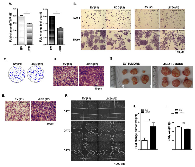
JAG1 expression is upregulated in high-grade metastatic prostate carcinomas and associated with poor disease-free survival of patients with prostate cancer. Intriguingly, all JAG1-positive prostate carcinomas express JICD although JICD function in prostate cancer (PC) cells is poorly understood. In this study, we found that JICD overexpression increased the expression levels of AR, especially AR-Vs, in PC cell lines and significantly enhanced androgen-independent and androgen-dependent function of ARs. Interestingly, JICD overexpression upregulated the expression of the PCSC marker CD133 in PC cells as the expression of self-renewal markers; namely, NANOG and OCT3/4 increased. In addition, JICD overexpression highly increased the expression of anti-apoptotic BCL-XL protein, while it little affected the expression of apoptotic BIM protein. In 3D cell culture assays, the spheres formed by JICD-overexpressing PC subline cells (C4-2 and CWR22Rv1) were larger than those formed by control (EV) subline cells with undifferentiated morphology. Although JICD overexpression caused quiescence in cell proliferation, it activated the expression of components in PCSC-related signaling pathways, increased PC cell mobility, and promoted in vivo xenograft mouse tumorigenesis. Therefore, JICD may play a crucial role in enhancing androgen independence and promoting stem-like properties in PC cells and should be considered a novel target for CRPC and PCSC diagnostic therapy.

Keywords: AR splicing variants; JAG1 intracellular domain; androgen receptor; androgen-independent activity; prostate cancer stem-like cells.


Conflict of interest statement

The authors declare no conflict of interest.

Figures

Figure 1
Figure 1
JAG1 regulates the expression of AR and AR–Vs. (A,B) Silencing of JAG1 decreases the expression of ARs. (A) Representative Western blot analysis showing the protein levels of AR and AR–Vs in siCtrl– or siJAG1–transfected androgen–dependent LNCaP and androgen-independent CWR22Rv1 cells. AR and AR–V proteins were detected using rabbit anti–AR (06–680, Millipore). (B) RT–PCR analysis showing the mRNA level of AR in siCtrl– or siJAG1–transfected LNCaP and CWR22Rv1 cells. RT–PCR analysis shows a decrease in total AR mRNAs, which contain exon 1–exon 2 (AR), in LNCaP and CWR22Rv1 cells when JAG1 was silenced. (C,D) Overexpression of JAG1 increases the expression of ARs. (C) Representative Western blot analysis showing the protein levels of AR and AR–Vs in EV– or JAG1–transfected C4–2 and CWR22Rv1 cells. AR and AR–V proteins were detected using rabbit anti–AR (06–680, Millipore). (D) RT–PCR analysis showing the level of AR mRNAs, which contain exon 3–exon 4 (AR), in EV or JAG1–transfected C4–2 and CWR22Rv1 cells. GAPDH was used as a loading control. Values above Western blots indicate the relative band intensity of each protein normalized to GAPDH and are shown as mean ± SD, which were obtained by densitometrical quantification of at least three independent experiments. All original uncropped Western blot and gel images are shown in Figure S7.
Figure 2
Figure 2
Processed intracellular domain of JAG1 (JICD) increases the expression of AR-Vs. (A) Representative Western blot analysis showing the increased protein levels of AR and AR–Vs in AdJICD–infected LNCaP and CWR22Rv1 cells compared to AdCtrl–infected cells. The proteins of AR and ARs including AR–V7 were detected using mouse anti-AR (sc–7305, Santa Cruz) and rabbit anti-AR–V7 (68492, Cell Signaling, Danvers, MA, USA) antibodies. (B,C) Stable JICD–overexpressing CWR22Rv1 subline cells show the increased expression of ARs, including AR–V7, at protein (B) and mRNA (C) levels. Representative RT–PCR (top) and quantitative RT–PCR (qPCR; bottom) analysis of three independent experiments (C) revealing a significant increase of AR–V7 mRNA expression in stable JICD–overexpressing CWR22Rv1 subline cells. GAPDH was used as a loading control. Data are shown as mean ± SEM, three independent experiments. *, p < 0.01; ns, not significant; two–tailed t-test. (D) JICD overexpression enriches the expression of genes involved in RNA processing and metabolic process, RNP complex biogenesis, and DNA metabolic process. (E) JICD upregulates the expression of some splicing factors involved in the generation of AR–V7 in CWR22Rv1 cells. (FH) JICD overexpression enhances the expression of some shared AR/AR–V7 (F) and AR–V7 target genes (G) and negatively affects the expression of most AR target genes (H). False discovery rate (FDR) < 0.05. Values above Western blots indicate the relative band intensity of each protein normalized to GAPDH and are shown as mean ± SD, which were obtained by densitometrical quantification of at least three independent experiments. All uncropped Western blot and gel original images are shown in Figure S7.
Figure 3
Figure 3
JAG1 and JICD enhance the androgen–dependent and androgen–independent transactivation of ARs. (AC) JICD overexpression significantly enhances the transactivation of ARs. Prostate cancer LNCaP (A), C4–2 (B), and CWR22Rv1 (C) cells were transiently transfected with FLAG–JICD or empty vector (EV) along with pARE2–TATA–luc and treated with 1 nM DHT or vehicle. (DF) PPC–1 cells overexpressed with AR–FL (D), AR–NTD–DBD (E), or AR–V7 (F) were transiently transfected with FLAG–JICD or empty vector (EV) and pARE2–TATA–luc and treated with 1 nM DHT or vehicle. (G) Overexpression of JAG1 and JICD enhances the N/C interaction of AR. PPC1 cells cotransfected with the AR N–terminal (VP16/AR1–660) domain, C–terminal (GAL4/AR–LBD658–919) domain, and 5xGAL4–luc3 reporter, along with JAG1, FLAG–JICD, or empty vector and incubated with 10 nM DHT. (H) Overexpression of JAG1 and JICD enhances coactivator recruitment to AR. PPC–1 cells were cotransfected with AR, SRC–1 (NCOA1), or SRC–2 (NCOA2), and JAG1, FLAG–JICD, or empty vector, along with pARE2–TATA–luc and incubated with 1 nM DHT. Luciferase activity was normalized to that of β-galactosidase. Data are shown as mean ± SEM. *, p < 0.05; **, p < 0.01; ***, p < 0.001; ns, not significant; one–way ANOVA with Tukey’s post–hoc test.
Figure 4
Figure 4
JICD promotes prostate cancer stem–like cell properties in prostate cancer cells. (A,B) Overexpression of JICD increases CSC marker expression in prostate cancer cells. Representative Western blot analysis (A) showing the protein levels of CD133, NANOG, BIM, BCL–XL, and JICD; Representative RT–PCR (top) and quantitative RT–PCR (qPCR; bottom) analysis of three independent experiments (B) revealing the mRNA levels of CD133 and JICD in stable EV– or JICD–overexpressing CWR22Rv1 subline cells. GAPDH was used as a loading control. Data are shown as mean ± SEM, three independent experiments. *, p < 0.01; two–tailed t-test. (C) Heatmap showing the strong upregulation of CD63, ASNS, TEX264, and FOXM1 and the downregulation of RB1 and P53 downstream signaling pathways (RB1, TP53BP2, TP53INP1, and RRM2B), which are related to stem-like cell properties. (D,E) JICD overexpression facilitates and maintains sphere formation. Representative images of stable EV– or JICD–overexpressing CWR22Rv1 spheres grown for the sphere formation (D, top) and modified 3D cell culture (E, top) assays (magnification, ×10 and ×20; bars, 400 and 200 μm, respectively). Arrowheads (D,E, top) indicate the spheres with differentiated morphology. Graphs presenting the number of spheres versus the diameter of spheres (μm) derived from stable EV– or JICD–overexpressing CWR22Rv1 cells, which were grown under sphere formation (D, bottom) and modified 3D cell culture (E, bottom) conditions. Data are shown as mean ± SEM. *, p < 0.05; **, p < 0.01; ns, not significant; one-way ANOVA with Tukey’s post-hoc test. (F) Representative Western blot analysis showing that spheres derived from stable JICD–overexpressing CWR22Rv1 cells expressed higher protein levels of CD133, NANOG, OCT3/4, and ARs, especially AR–Vs, including AR–V7, than the control EV cell-derived spheres did. Values above Western blots indicate the relative band intensity of each protein normalized to GAPDH and are shown as mean ± SD, which were obtained by densitometrical quantification of at least three independent experiments. All original uncropped Western blot and gel images are shown in Figure S7.
Figure 5
Figure 5
JICD overexpression increases prostate cancer cell mobility and in vivo tumorigenesis. (A,B) Stable JICD overexpression reduces the proliferation and viability of CWR22Rv1 cells. Stable EV (#1) or JICD (#2) CWR22Rv1 subline cells were maintained for 3 days and then subjected to MTS/PMS assays (A, left) or cell counting assay (A, right). Stable EV (#1 and #3) or JICD (#2 and #4) subline cells were maintained for 1 and 6 days, stained with crystal violet, and imaged via ZEISS microscopy at 20× magnification (scale bar 10 μm) (B). Data are shown as mean ± SEM. *, p < 0.01; two-tailed t-test. (C) Colony size increases in stable JICD-overexpressing CWR22Rv1 subline cells compared with that in the control without significant difference in the total number of colonies between them. Stable EV (#1) or JICD (#2) subline cells were maintained for 2 weeks and then processed for 0.5% crystal violet staining. Colonies were imaged with a normal camera. (DF) JICD increases the mobility of prostate cancer cells. Invasion assays of stable EV (#1) or JICD (#2) CWR22Rv1 subline cells (D). Cell invasion was allowed to occur for 48 h, stained with crystal violet, and imaged via ZEISS microscopy at 10× magnification (Scale bar = 10 μm). Boyden Chamber migration assay of stable EV– or JICD–overexpressing CWR22Rv1 cells (E). Cells were allowed to migrate for 24 h, stained with crystal violet, and imaged with ZEISS microscopy at 10× magnification (Scale bar = 10 μm). Scratch wound-closure assay of stable EV (#1) or JICD (#2) CWR22Rv1 subline cells (F). Scratch wounds were generated (day 0, D0), and wound distances were monitored for 2 (D2) and 4 (D4) days; they were then imaged using EVOS® FL Cell Imaging System at 4× magnification (Scale bar = 1000 μm). Boundary lines were simply drawn using the shape tool from PowerPoint. (GI) JICD overexpression promotes CWR22Rv1 xenograft tumor growth in vivo. Stable JICD–overexpressing (JICD; #2) or control (EV; #1) CWR22Rv1 subline cells were implanted into the shoulders of 4–week–old male NOD.CB17–PrkdcSCID/J mice, and tumors were allowed to grow for 7 weeks. Representative tumors (G) dissected from mice after 7 weeks. Tumor weight (H) and body weight (I) measured after 7 weeks. Data are shown as mean ± SEM, two independent biological in vivo experiments (n = 6 mice per each group). *, p < 0.01; ns, not significant; two–tailed t-test.

Similar articles

Cited by

References

    1. Tan M.H., Li J., Xu H.E., Melcher K., Yong E.L. Androgen receptor: Structure, role in prostate cancer and drug discovery. Acta Pharmacol. Sin. 2015;36:3–23. doi: 10.1038/aps.2014.18. - DOI - PMC - PubMed
    1. Sharifi N. Mechanisms of androgen receptor activation in castration-resistant prostate cancer. Endocrinology. 2013;154:4010–4017. doi: 10.1210/en.2013-1466. - DOI - PMC - PubMed
    1. Dai C., Heemers H., Sharifi N. Androgen Signaling in Prostate Cancer. Cold Spring Harb. Perspect. Med. 2017;7:a030452. doi: 10.1101/cshperspect.a030452. - DOI - PMC - PubMed
    1. Wadosky K.M., Koochekpour S. Androgen receptor splice variants and prostate cancer: From bench to bedside. Oncotarget. 2017;8:18550–18576. doi: 10.18632/oncotarget.14537. - DOI - PMC - PubMed
    1. Sprenger C.C., Plymate S.R. The link between androgen receptor splice variants and castration-resistant prostate cancer. Horm. Cancer. 2014;5:207–217. doi: 10.1007/s12672-014-0177-y. - DOI - PMC - PubMed