Review
. 2022 Nov 16;12(11):2810.
doi: 10.3390/diagnostics12112810.

Placental and Umbilical Cord Anomalies Diagnosed by Two- and Three-Dimensional Ultrasound

Affiliations
Review

Placental and Umbilical Cord Anomalies Diagnosed by Two- and Three-Dimensional Ultrasound

Eduardo Félix Martins Santana et al. Diagnostics (Basel). .

Abstract

The aim of this review is to present a wide spectrum of placental and umbilical cord pathologies affecting the pregnancy. Placental and umbilical cord anomalies are highly associated with high-risk pregnancies and may jeopardize fetal well-being in utero as well as causing a predisposition towards poor perinatal outcome with increased fetal and neonatal mortality and morbidity. The permanent, computerized perinatology databases of different international centers have been searched and investigated to fulfil the aim of this manuscript. An extended gallery of prenatal imaging with autopsy correlation in specific cases will help to provide readers with a useful iconographic tool and will assist with the understanding and definition of this critical obstetrical and perinatological issue.

Keywords: autopsy; perinatal outcome; placental pathology; three-dimensional ultrasound; two-dimensional ultrasound; umbilical cord pathology.


Conflict of interest statement

The authors declare no conflict of interest.

Figures

Figure 1
Figure 1
Two-dimensional ultrasound imaging showing PAS (placenta accreta spectrum, placenta increta type): longitudinal scan in a patient at 24 week’s gestation shows multiple hypoechoic areas (lacunae) (white arrows). Color Doppler ultrasound shows increased vascularity and the bulging of the placenta increta (red arrow); (bl): bladder.
Figure 2
Figure 2
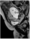
Fetal MRI (13 weeks, sagittal T2) showing an early diagnosis in a case of PAS (placenta accreta, type) (Legend: AF, amniotic fluid; Bl, bladder; Pl, placenta).
Figure 3
Figure 3
Transvaginal two-dimensional ultrasound showing placenta praevia major (upper panel, a,b) and a placenta praevia minor (lower panel, c,d).
Figure 4
Figure 4
Fetal MRI (sagittal T2) performed at 30 weeks and 26 week’s gestation in two different fetuses (A,B), demonstrating a placenta praevia major and accretism. (Legend: Pl: placenta; Cx: cervix).
Figure 5
Figure 5
Two-dimensional color Doppler ultrasound and three-dimensional ultrasound using HD-Live™ flow showing a bilobed placenta. When an accessory lobe is detected (succenturiate) in the lower part of the uterus, careful evaluation with ultrasound for vasa praevia and velamentous cord insertion should be performed.
Figure 6
Figure 6
Pregnancy at 20 week’s gestation. Two-dimensional color Doppler ultrasound showing velamentous insertion in a bilobed placenta. (Legend: UA, umbilical artery).
Figure 7
Figure 7
Pregnancy at 21 weeks’ gestation. Succenturiate placenta and placenta praevia Type II. Color Doppler and four-dimensional (4D) STIC with color Doppler. Low-lying placenta and accessory lobe are connected by intramembranous blood vessels, with the umbilical cord originating from the main placenta (A). Vasa praevia and contemporaneous cord procidentia are highlighted (cervical canal is indicated by a small arrow) (B). Pathologic correlation (C,D) showed two separate placental lobes interconnected by membranes, with blood vessels traversing the membranes (indicated by curved arrows).
Figure 8
Figure 8
Circumvallate placenta with anterior placenta at two-dimensional ultrasound (A). Three-dimensional ultrasound using HD-Live Silhouette™ mode (B) at 20 week’s gestation shows the raised edge of the placenta as a linear band of tissue that may mimic a uterine synaechia (arrows).
Figure 9
Figure 9
(A,B) Circumvallate placenta (pl.) with posterior placenta. Two-dimensional and three-dimensional ultrasound using HD-Live™ mode showing a “shelf-like” structure.
Figure 10
Figure 10
(A,B) Fetal MRI (coronal T1 and T2) in a case of circumvallate placenta at 28 week’s gestation (white arrows).
Figure 11
Figure 11
Placental with mesenchymal dysplasia at 15 weeks of gestation. Two-dimensional (A,B) and three-dimensional ultrasound (C,D) with HD-Live Silhouette™ and inversion mode displaying a characteristic large placenta with “grape-like”, multicystic changes.
Figure 12
Figure 12
Three-dimensional ultrasound in a case of placental chorioangioma detected using Cristal Vue™ at 31 week’s gestation (A). Note the detail of the vascular branching (B).
Figure 13
Figure 13
Chorioangioma. Pregnancy at 20 and 26 week’s gestation. Two-dimensional color Doppler (A), three-dimensional ultrasound with HD-live™ surface rendering (B) and with Silhouette™ effect (C). Chorioangioma arises from the fetal surface of the placenta, where vessels’ cords diverge before joining the placenta.
Figure 14
Figure 14
Two-dimensional (A,B) and three-dimensional ultrasound with HD flow using “glass body” mode (C) showing chorionic plate cyst. It depicts an anechoic thin-walled structure on the fetal surface of the placenta near the cord insertion (arrows) (A,B) without internal vascularity (C). Pathologic correlation (arrows) (D).
Figure 15
Figure 15
Two-dimensional ultrasound using color Doppler ultrasound and three-dimensional ultrasound with STIC color Doppler and “glass body” mode rendering with HD-Live™ flow showing multiple loops of cord around the neck in sagittal (A), axial (B) and coronal scans (C), resembling a “whirlpool” sign.
Figure 16
Figure 16
Second trimester transvaginal scan and three-dimensional ultrasound with “glass-body” rendering (A), Doppler ultrasound (B) and two-dimensional ultrasound showing the anatomical relationship between the cervical canal (yellow lines) (C) and the presence of vasa praevia (red arrow) (D).
Figure 17
Figure 17
Pregnancy at 24 weeks’ gestation. Two-dimensional ultrasound, color and pulse wave (PW) spectral Doppler ultrasound allowed the detection of an arterial vessel close to the internal cervical os (vasa praevia Type III, white arrows)(Legend: CC: cervical canal).
Figure 18
Figure 18
(A,B) First trimester (12 weeks) scan showing in two-dimensional- and three-dimensional color Doppler ultrasound a velamentous cord insertion (Cordone: umbilical cord).
Figure 19
Figure 19
Pregnancy at 19 week’s gestation (same case of the previous figure). Two-dimensional color Doppler ultrasound (A) and four-dimensional color Doppler STIC volume (B) diagnosed velamentous cord insertion.
Figure 20
Figure 20
Velamentous cord insertion into the placental membranes (A) with direction of blood flow indicated by curved arrows and pathologic correlation (arrows) (B).
Figure 21
Figure 21
Twin pregnancy at 16 week’s gestation. Two-dimensional ultrasound (A) and four-dimensional STIC with color Doppler ultrasound (B) shows a particular velamentous insertion of the cord’s vessels that run through the intertwin membranes before joining the placenta (arrows).
Figure 22
Figure 22
Pregnancy at 20 weeks gestation. Two-dimensional ultrasound (A), color Doppler (B) and four-dimensional STIC with color Doppler ultrasound (C). In velamentous insertion, the umbilical cord vessels (UC) diverge from each other, inserting into the membranes not supported by Wharton jelly, before entering the placental tissue (curved arrows, figure on the left) (A). The diagnosis of velamentous cord insertion is made through color Doppler ultrasound by the discovery of splayed umbilical vessels at the periphery of the placenta (B). It may be associated with vasa praevia (Type I) when umbilical vessels running through the fetal membranes are close to the internal cervical os (figure on the center). Four-dimensional STIC with color Doppler ultrasound (C) image showing the “mangrove sign” for velamentous cord insertion.
Figure 23
Figure 23
Marginal insertion of the umbilical cord using HD-Flow™ Doppler ultrasound.
Figure 24
Figure 24
Furcate umbilical cord insertion using two-dimensional ultrasound (A) and three-dimensional ultrasound (B) with HD-Flow™ Doppler: the dichotomic direction of the umbilical blood flow is clearly demonstrated at 17.3 week’s gestation (arrows).
Figure 25
Figure 25
MCMA twin pregnancy. Cord entanglement can also be detected with the “galloping horse sign” that involves identifying two distinct waves signals with different heart rates obtained by sampling an apparently unique umbilical cord with pulse wave (PW) spectral Doppler ultrasound.
Figure 26
Figure 26
Sonographic evaluation of umbilical cord hemangioma. (Legend: AO: umbilical artery; VO: umbilical vein). Umbilical cord hemangioma or angiomyxoma is usually located in the terminal portion of the cord on the placental side. Two-dimensional color (A) and power Doppler ultrasound (B) may detect a fusiform or saccular swelling of the umbilical cord. Umbilical arteries are surrounded by a particularly echogenic material arranged as a sheath of the same vessels. The mechanical compression of the umbilical circulation may be determined by an enlargement of the mass and stenosis of the umbilical vessels also caused by the intraluminal proliferation of the hemangioma. Blood flow reduction can be abruptly compromised through the twisting of the cord favored by the mass and by the presence of pseudocysts. An accurate follow-up is mandatory.
Figure 27
Figure 27
Pathologic correlation. Umbilical cord hemangioma at the fetal face of the placenta (A) and the axial section of the umbilical cord (B) showing a large hematoma in one of the two umbilical artery (UA: umbilical artery; UV: umbilical vein). Evident increase in the diameter of the cord due to swelling and myxoid-edematous degeneration. The section of the cord shows the two umbilical arteries surrounded by thick angiomatous tissue.
Figure 28
Figure 28
Transient umbilical cord hematoma in the cord insertion area on the placenta after intra uterine fetal transfusion (IUFT). Note the absence of blood flow on color Doppler ultrasound assessment (arrows).
Figure 29
Figure 29
Pregnancy at 12 week’s gestation. Two-dimensional ultrasound: sagittal scan of the fetus showing a cyst filled with finely corpuscular material at the level of the cord insertion on fetal abdomen (A). Color Doppler ultrasound highlights the cord’s splayed vessels due to a hemorrhagic cyst with a clot inside it (arrow) (B). (Legend: Pl: placenta).
Figure 30
Figure 30
Doppler ultrasound showing a single umbilical artery (SUA) coursing along the bladder.
Figure 31
Figure 31
Pregnancy at 22 week’s gestation. Color Doppler (AC) and four-dimensional STIC with color Doppler ultrasound in “glass-body” mode (D). Longitudinal and axial scan of the cord (AC) demonstrate the presence of a single umbilical artery (SUA). SUA shows a particular paravesical kinking (Bl: bladder).
Figure 32
Figure 32
Doppler ultrasound (A) demonstrating an umbilical cord varix (curved arrows) with pathology confirmation after birth (B).
Figure 33
Figure 33
Pregnancy at 32, 34 and 35 week’s gestation. Two-dimensional ultrasound in longitudinal and axial scan of the umbilical cord. A moderate echogenicity corresponds to segmental thrombosis of an umbilical artery. The thrombosis zone is indicated by red arrows and the progressive extension is further delimited by arrows (Legend: A: umbilical artery; V: umbilical vein).
Figure 34
Figure 34
Sonographic view of umbilical cord vessel aneurism with color Doppler ultrasound (A). Gross anatomy: umbilical cord after delivery confirmed presence of multiples aneurisms (B).

References

    1. Rezende G.C., Araujo E., Jr. Prenatal diagnosis of placenta and umbilical cord pathologies by three-dimensional ultrasound: Pictorial essay. Med. Ultrason. 2015;14:545–549. - PubMed
    1. Ptacek I., Sebire N.J., Man J.A., Brownbill P., Heazell A.E.P. Systematic review of placental pathology reported in association with stillbirth. Placenta. 2014;5:552–562. - PubMed
    1. Bukowski R., Hansen N.I., Pinar H., Willinger M., Reddy U.M., Parker C.B., Silver R.M., Dudley D.J., Stoll B.J., Saade G.R., et al. Altered fetal growth, placental abnormalities, and stillbirth. PLoS ONE. 2017;12:e0182874. doi: 10.1371/journal.pone.0182874. - DOI - PMC - PubMed
    1. Hayes D.J.L., Warland J., Parast M.M., Bendon R.W., Hasegawa J., Banks J., Clapham L., Heazell A.E.P. Umbilical cord characteristics and their association with adverse pregnancy outcomes: A systematic review and meta-analysis. PLoS ONE. 2020;15:e0239630. doi: 10.1371/journal.pone.0239630. - DOI - PMC - PubMed
    1. Hammad I.A., Blue N.R., Allshouse A.A., Silver R.M., Gibbins K.J., Page J.M., Goldenberg R.L., Reddy U.M., Saade G.R., Dudley D.J., et al. Umbilical Cord Abnormalities and Stillbirth. Obstet. Gynecol. 2020;135:644–652. doi: 10.1097/AOG.0000000000003676. - DOI - PMC - PubMed

LinkOut - more resources