

The Use of CBCT in Evaluating the Health and Pathology of the Maxillary Sinus
- PMID: 36428879
- PMCID: PMC9689855
- DOI: 10.3390/diagnostics12112819
The Use of CBCT in Evaluating the Health and Pathology of the Maxillary Sinus
Abstract
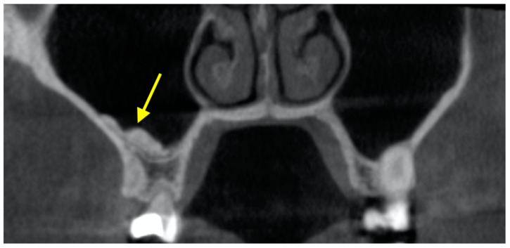
The use of cone-beam computed tomography (CBCT) has been increasing in dental practice. This narrative review summarized the relevance and utilizations of CBCT to visualize anatomical structures of the maxillary sinus and common pathologies found in the maxillary sinus. The detection/visualization rate, the location and the morphometric characteristics were described. For sinus anatomy, the reviewed features included the posterior superior alveolar artery, sinus pneumatization, sinus hypoplasia, sinus septa, and primary and accessory sinus ostia. For pathology, the following items were reviewed: membrane thickening associated with periapical lesions/periodontal lesions, mucous retention cyst, and antrolith. The visualization and assessment of the maxillary sinus is very important prior to procedures that take place in close proximity with the sinus floor, such as tooth extraction, implant insertion, and sinus floor elevation. Some sinus pathologies may be associated with odontogenic lesions, such as periapical diseases and periodontal bone loss.
Keywords: CBCT; Schneiderian membrane; cone beam computed tomography; health; maxillary sinus; pathology; sinus floor elevation.
Conflict of interest statement
The authors declare no conflict of interest.
Figures

Similar articles
-
Evaluation of Association between Maxillary Posterior Teeth Periapical Pathologies and Maxillary Sinus Mucosal Changes-A Cone-Beam Computed Tomography (CBCT) Study.Indian J Radiol Imaging. 2023 Nov 23;34(2):246-253. doi: 10.1055/s-0043-1777013. eCollection 2024 Apr. Indian J Radiol Imaging. 2023. PMID: 38549905 Free PMC article.
-
The Impact of Posterior Maxillary Teeth on Maxillary Sinus: Insights From Cone-Beam Computed Tomography Analysis.Cureus. 2024 Dec 29;16(12):e76578. doi: 10.7759/cureus.76578. eCollection 2024 Dec. Cureus. 2024. PMID: 39877792 Free PMC article.
-
The relation between Schneiderian membrane thickening and radiodiagnostic features of periapical pathology.Braz Dent J. 2024 Sep 16;35:e245775. doi: 10.1590/0103-6440202405775. eCollection 2024. Braz Dent J. 2024. PMID: 39320000 Free PMC article.
-
Open Sinus Lift Surgery and the Importance of Preoperative Cone-Beam Computed Tomography Scan: A Review.J Int Oral Health. 2015 Sep;7(9):127-33. J Int Oral Health. 2015. PMID: 26435632 Free PMC article. Review.
-
CBCT for Diagnostics, Treatment Planning and Monitoring of Sinus Floor Elevation Procedures.Diagnostics (Basel). 2023 May 10;13(10):1684. doi: 10.3390/diagnostics13101684. Diagnostics (Basel). 2023. PMID: 37238169 Free PMC article. Review.
Cited by
-
A platform combining automatic segmentation and automatic measurement of the maxillary sinus and adjacent structures.Clin Oral Investig. 2025 Jan 25;29(1):88. doi: 10.1007/s00784-025-06191-x. Clin Oral Investig. 2025. PMID: 39862338
-
A Cone Bean Computer Tomography Investigation of the Newly Formed Mandibular Anterior Ridge following the Treatment of an Extended Comminuted Fracture: A 12-Year Follow-Up.Case Rep Dent. 2024 Feb 21;2024:1824016. doi: 10.1155/2024/1824016. eCollection 2024. Case Rep Dent. 2024. PMID: 38419613 Free PMC article.
-
The Impact of Maxillary Sinus Pneumatization on the Quality of the Alveolar Bone in Dentated and Edentulous Patients: A Cone-Beam Computed Tomography Radiographic Analysis.Cureus. 2023 Sep 26;15(9):e46005. doi: 10.7759/cureus.46005. eCollection 2023 Sep. Cureus. 2023. PMID: 37900530 Free PMC article.
-
Application of Cone Beam Computed Tomography in Risk Assessment of Lower Third Molar Surgery.Diagnostics (Basel). 2023 Mar 1;13(5):919. doi: 10.3390/diagnostics13050919. Diagnostics (Basel). 2023. PMID: 36900063 Free PMC article. Review.
-
Current Applications of Deep Learning and Radiomics on CT and CBCT for Maxillofacial Diseases.Diagnostics (Basel). 2022 Dec 29;13(1):110. doi: 10.3390/diagnostics13010110. Diagnostics (Basel). 2022. PMID: 36611402 Free PMC article. Review.
References
-
- Braun M.J., Rauneker T., Dreyhaupt J., Hoffmann T.K., Luthardt R.G., Schmitz B., Dammann F., Beer M. Dental and Maxillofacial Cone Beam CT—High Number of Incidental Findings and Their Impact on Follow-Up and Therapy Management. Diagnostics. 2022;12:1036. doi: 10.3390/diagnostics12051036. - DOI - PMC - PubMed
-
- Kirkham-Ali K., La M., Sher J., Sholapurkar A. Comparison of cone-beam computed tomography and panoramic imaging in assessing the relationship between posterior maxillary tooth roots and the maxillary sinus: A systematic review. J. Investig. Clin. Dent. 2019;10:e12402. doi: 10.1111/jicd.12402. - DOI - PubMed
Publication types
LinkOut - more resources
Full Text Sources
