Review
. 2022 Nov 16;12(11):2823.
doi: 10.3390/diagnostics12112823.

Challenges in the Differential Diagnosis of COVID-19 Pneumonia: A Pictorial Review

Affiliations
Review

Challenges in the Differential Diagnosis of COVID-19 Pneumonia: A Pictorial Review

Cristina Maria Marginean et al. Diagnostics (Basel). .

Abstract

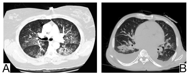
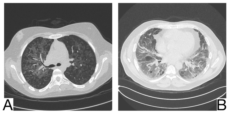
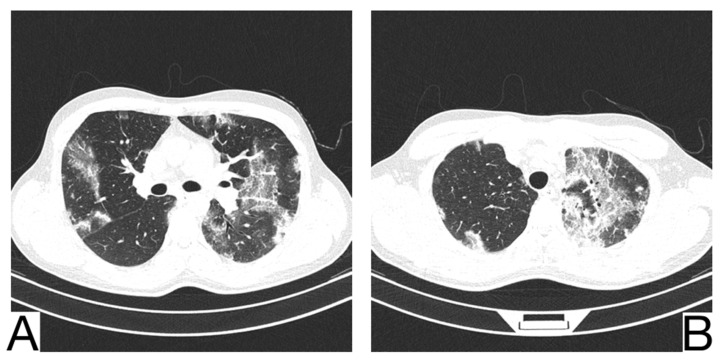
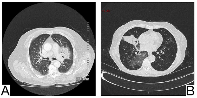
COVID-19 pneumonia represents a maximum medical challenge due to the virus's high contagiousness, morbidity, and mortality and the still limited possibilities of the health systems. The literature has primarily focused on the diagnosis, clinical-radiological aspects of COVID-19 pneumonia, and the most common possible differential diagnoses. Still, few studies have investigated the rare differential diagnoses of COVID-19 pneumonia or its overlap with other pre-existing lung pathologies. This article presents the main radiological features of COVID-19 pneumonia and the most common alternative diagnoses to establish the vital radiological criteria for a differential diagnosis between COVID-19 pneumonia and other lung pathologies with similar imaging appearance. The differential diagnosis of COVID-19 pneumonia is challenging because there may be standard radiologic features such as ground-glass opacities, crazy paving patterns, and consolidations. A multidisciplinary approach is crucial to define a correct final diagnosis, as an overlap of COVID-19 pneumonia with pre-existing lung diseases is often possible and suggests possible differential diagnoses. An optimal evaluation of HRTC can help limit the clinical evolution of the disease, promote therapy for patients and ensure an efficient allocation of human and economic resources.

Keywords: COVID-19; differential diagnosis; pneumonia.


Conflict of interest statement

The authors declare no conflict of interest.

Figures

Figure 1
Figure 1
Symptomatology of COVID-19 (based on data published by Guan et al. [13] and Zhou et al. [11]).
Figure 2
Figure 2
Main comorbidities in COVID-19 patients (based on data published by Guan et al. [13] and Zhou et al. [11]).
Figure 3
Figure 3
Laboratory findings in COVID-19 patients (based on data published by Guan et al. [13] and Zhou et al. [11]).
Figure 4
Figure 4
Imagistic findings in COVID-19 (A). Multiple areas of ground glass infiltration (patient on the third day of symptoms) (B). Bilateral patches of ground glass and subsegmental consolidation (C). Ground glass and consolidation with air bronchogram (8 days after onset) (D). Diffuse ground glass infiltration (white lung appearance). Note. Adapted from Hefeda et al. (2020) [22].
Figure 5
Figure 5
(A). Consolidation with air bronchogram in a patient with bacterial pneumonia (B). Consolidation and bronchovascular bundle thickening in a patient with Mycoplasma pneumoniae pneumonia. Note. Adapted from Mikael Häggström, M.D (A) and Tanaka (2016) (B).
Figure 6
Figure 6
(A). Multifocal areas of poorly defined focal consolidation in a patient positive for influenza A (H1N1) (B). Bilateral ground-glass opacities and dense airspace consolidations in a patient with Pneumocystis jirovecii pneumonia (C). Halo sign in a patient with aspergillosis. Note. Adapted from Elmokadem et al. (2021) [40] (A), Sullivan et al. [41] (2020) (B), and Dr. Laughlin Dawes (C).
Figure 7
Figure 7
Acute pulmonary edema (A). Ground glass opacity in mainly perihilar and dependent distribution (B). Bilateral airspace opacification in central peribronchovascular distribution and smooth interlobular septal thickening (indicating interstitial edema) and moderate bilateral pleural effusion. Note. Case courtesy of The Radswiki, Radiopaedia.org, rID: 1183 (A) and Dr. Rania Adel Anan, Radiopaedia.org, rID: 95825 (B).
Figure 8
Figure 8
Diffuse alveolar hemorrhage, pulmonary parenchyma with a diffuse increase in density, and bilateral alveolar filling pattern, predominantly in lower lobes (A,B). Note—case courtesy of Dr. Jesus Sanchez Castro, Radiopaedia.org, rID: 68769.
Figure 9
Figure 9
Hypersensitivity pneumonia (A). Perihilar ground glass changes (B). Gas trapping on the expiratory image, ground glass opacity, and honeycombing. Note. Case courtesy of Dr. Yi-Jin Kuok, Radiopaedia.org, rID: 17192 (A) and Dr. Henry Knipe, Radiopaedia.org, rID: 48107 (B).
Figure 10
Figure 10
Chronic eosinophilic pneumonia (A,B). Consolidation throughout both lungs. Note. Case courtesy of Dr. Henry Knipe, Radiopaedia.org, rID: 39331.
Figure 11
Figure 11
Lipoid pneumonia (A). Ill-defined airspace opacity next to the left hilum (B). Atelectasis of middle lobe and ground-glass opacity. Note. Case courtesy of Dr. Aneta Kecler-Pietrzyk, Radiopaedia.org, rID: 62113 (A) and Dr. Abraão Kupske, Radiopaedia.org, rID: 55752 (B).
Figure 12
Figure 12
Pulmonary alveolar proteinosis (A,B). Bilateral areas of crazy paving. Note. Case courtesy of Dr. Adrià Roset Altadill, Radiopaedia.org, rID: 74896.
Figure 13
Figure 13
Idiopathic interstitial pneumonia (A,B). Bilateral septal thickening and honeycombing with more severe involvement toward the lung bases. Note. Case courtesy of Dr. Hani Makky Al Salam, Radiopaedia.org, rID: 41974.
Figure 14
Figure 14
Pulmonary infarction (A,B). Wedge-shaped peripheral consolidation, absent air bronchogram. Note. Case courtesy of Dr. Vladislav Boyanov Rusinov, Radiopaedia.org, rID: 19479 (A) and Assoc Prof Craig Hacking, Radiopaedia.org, rID: 73062 (B).

Similar articles

Cited by

References

    1. Cottin V., Cordier J.-F. Eosinophilic pneumonias. Allergy. 2005;60:841–857. doi: 10.1111/j.1398-9995.2005.00812.x. - DOI - PubMed
    1. Pollard C.A., Morran M.P., Nestor-Kalinoski A.L. The COVID-19 pandemic: A global health crisis. Physiol. Genom. 2020;52:549–557. doi: 10.1152/physiolgenomics.00089.2020. - DOI - PMC - PubMed
    1. Eastin C., Eastin T. Clinical Characteristics of Coronavirus Disease 2019 in China. J. Emerg. Med. 2020;58:711–712. doi: 10.1016/j.jemermed.2020.04.004. - DOI
    1. Sharma R., Agarwal M., Gupta M., Somendra S., Saxena S.K. Coronavirus Disease 2019 (COVID-19) Springer; Singapore: 2020. Clinical Characteristics and Differential Clinical Diagnosis of Novel Coronavirus Disease 2019 (COVID-19) pp. 55–70.
    1. Wu Z., McGoogan J.M. Characteristics of and Important Lessons from the Coronavirus Disease 2019 (COVID-19) Outbreak in China: Summary of a Report of 72,314 Cases from the Chinese Center for Disease Control and Prevention. JAMA. 2020;323:1239–1242. doi: 10.1001/jama.2020.2648. - DOI - PubMed

LinkOut - more resources