Claudin-10 Expression Is Increased in Endometriosis and Adenomyosis and Mislocalized in Ectopic Endometriosis
- PMID: 36428908
- PMCID: PMC9689821
- DOI: 10.3390/diagnostics12112848
Claudin-10 Expression Is Increased in Endometriosis and Adenomyosis and Mislocalized in Ectopic Endometriosis
Abstract
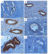
Claudins, as the major components of tight junctions, are crucial for epithelial cell-to-cell contacts. Recently, we showed that in endometriosis, the endometrial epithelial phenotype is highly conserved, with only minor alterations. For example, claudin-11 is strongly expressed; however, its localization in the endometriotic epithelial cells was impaired. In order to better understand the role of claudins in endometrial cell-to-cell contacts, we analyzed the tissue expression and localization of claudin-10 by immunohistochemistry analysis and two scoring systems. We used human tissue samples (n = 151) from the endometrium, endometriosis, and adenomyosis. We found a high abundance of claudin-10 in nearly all the endometrial (98%), endometriotic (98−99%), and adenomyotic (90−97%) glands, but no cycle-specific differences and no differences in the claudin-10 positive endometrial glands between cases with and without endometriosis. A significantly higher expression of claudin-10 was evident in the ectopic endometrium of deep-infiltrating (p < 0.01) and ovarian endometriosis (p < 0.001) and in adenomyosis in the cases with endometriosis (p ≤ 0.05). Interestingly, we observed a shift in claudin-10 from a predominant apical localization in the eutopic endometrium to a more pronounced basal/cytoplasmic localization in the ectopic endometria of all three endometriotic entities but not in adenomyosis. Significantly, despite the impaired endometriotic localization of claudin-10, the epithelial phenotype was retained. The significant differences in claudin-10 localization between the three endometriotic entities and adenomyosis, in conjunction with endometriosis, suggest that most of the aberrations occur after implantation and not before. The high similarity between the claudin-10 patterns in the eutopic endometrial and adenomyotic glands supports our recent conclusions that the endometrium is the main source of endometriosis and adenomyosis.
Keywords: adenomyosis; claudin-10; endometriosis; endometrium.
Conflict of interest statement
The author(s) declare no potential conflict of interest with respect to the research, authorship, and/or publication of this article.
Figures


Similar articles
-
The Different Gene Expression Profile in the Eutopic and Ectopic Endometrium Sheds New Light on the Endometrial Seed in Endometriosis.Biomedicines. 2024 Jun 8;12(6):1276. doi: 10.3390/biomedicines12061276. Biomedicines. 2024. PMID: 38927483 Free PMC article. Review.
-
Impaired Localization of Claudin-11 in Endometriotic Epithelial Cells Compared to Endometrial Cells.Reprod Sci. 2019 Sep;26(9):1181-1192. doi: 10.1177/1933719118811643. Epub 2018 Dec 4. Reprod Sci. 2019. PMID: 30514158
-
Localization of claudin-2 and claudin-3 in eutopic and ectopic endometrium is highly similar.Arch Gynecol Obstet. 2020 Apr;301(4):1003-1011. doi: 10.1007/s00404-020-05472-y. Epub 2020 Mar 5. Arch Gynecol Obstet. 2020. PMID: 32140805 Free PMC article.
-
Impaired Expression of Membrane Type-2 and Type-3 Matrix Metalloproteinases in Endometriosis but Not in Adenomyosis.Diagnostics (Basel). 2022 Mar 22;12(4):779. doi: 10.3390/diagnostics12040779. Diagnostics (Basel). 2022. PMID: 35453827 Free PMC article.
-
Adenomyosis pathogenesis: insights from next-generation sequencing.Hum Reprod Update. 2021 Oct 18;27(6):1086-1097. doi: 10.1093/humupd/dmab017. Hum Reprod Update. 2021. PMID: 34131719 Free PMC article. Review.
Cited by
-
Neutrophil gelatinase-associated lipocalin serum level: A potential noninvasive biomarker of endometriosis?Medicine (Baltimore). 2023 Oct 13;102(41):e35539. doi: 10.1097/MD.0000000000035539. Medicine (Baltimore). 2023. PMID: 37832065 Free PMC article.
-
How Do Microorganisms Influence the Development of Endometriosis? Participation of Genital, Intestinal and Oral Microbiota in Metabolic Regulation and Immunopathogenesis of Endometriosis.Int J Mol Sci. 2023 Jun 30;24(13):10920. doi: 10.3390/ijms241310920. Int J Mol Sci. 2023. PMID: 37446108 Free PMC article. Review.
-
The Different Gene Expression Profile in the Eutopic and Ectopic Endometrium Sheds New Light on the Endometrial Seed in Endometriosis.Biomedicines. 2024 Jun 8;12(6):1276. doi: 10.3390/biomedicines12061276. Biomedicines. 2024. PMID: 38927483 Free PMC article. Review.
-
Causal association between telomere length and female reproductive endocrine diseases: a univariable and multivariable Mendelian randomization analysis.J Ovarian Res. 2024 Jul 15;17(1):146. doi: 10.1186/s13048-024-01466-5. J Ovarian Res. 2024. PMID: 39010148 Free PMC article.
References
-
- Sampson J.A. Peritoneal endometriosis due to menstrual dissemination of endometrial tissue into the peritoneal cavity. Am. J. Obstet. Gynecol. 1927;14:422–469. doi: 10.1016/S0002-9378(15)30003-X. - DOI
-
- Blumenkrantz M.J., Gallagher N., Bashore R.A., Tenckhoff H. Retrograde menstruation in women undergoing chronic peritoneal dialysis. Obstet. Gynecol. 1981;57:667–670. - PubMed
-
- Halme J., Hammond M.G., Hulka J.F., Raj S.G., Talbert L.M. Retrograde menstruation in healthy women and in patients with endometriosis. Obstet. Gynecol. 1984;64:151–154. - PubMed
LinkOut - more resources
Full Text Sources

