Review
. 2022 Nov 21;11(22):3698.
doi: 10.3390/cells11223698.

The Role of IL-6 in Cancer Cell Invasiveness and Metastasis-Overview and Therapeutic Opportunities

Affiliations
Review

The Role of IL-6 in Cancer Cell Invasiveness and Metastasis-Overview and Therapeutic Opportunities

Magdalena Rašková et al. Cells. .

Abstract

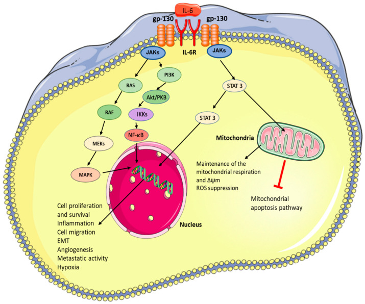
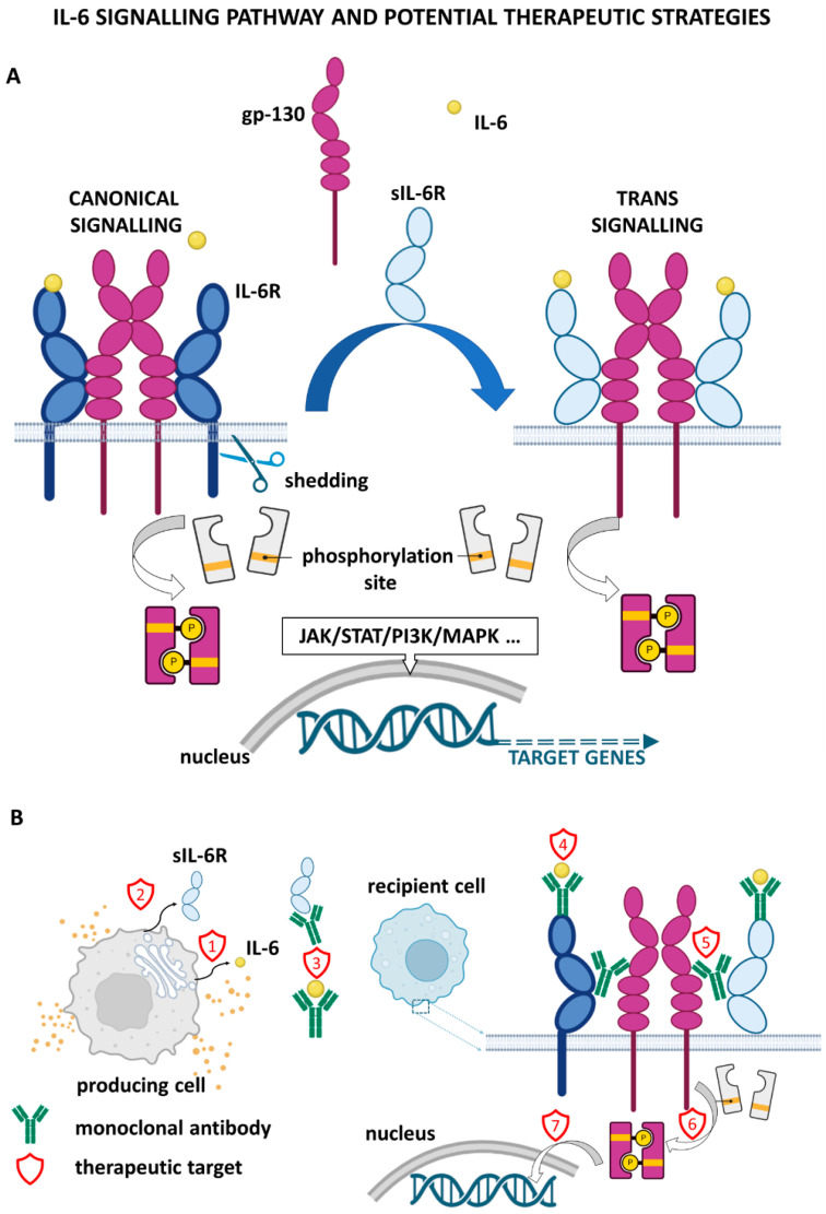
Interleukin 6 (IL-6) belongs to a broad class of cytokines involved in the regulation of various homeostatic and pathological processes. These activities range from regulating embryonic development, wound healing and ageing, inflammation, and immunity, including COVID-19. In this review, we summarise the role of IL-6 signalling pathways in cancer biology, with particular emphasis on cancer cell invasiveness and metastasis formation. Targeting principal components of IL-6 signalling (e.g., IL-6Rs, gp130, STAT3, NF-κB) is an intensively studied approach in preclinical cancer research. It is of significant translational potential; numerous studies strongly imply the remarkable potential of IL-6 signalling inhibitors, especially in metastasis suppression.

Keywords: IL-6; cancer; metastasis.


Conflict of interest statement

The authors declare no conflict of interest.

Figures

Figure 1
Figure 1
Expression of the IL6 gene in stromal elements of melanoma and head and neck squamous cell carcinoma (HNSCC). Normal dermal fibroblasts (DF, (left panel)) and normal mucous fibroblasts (MuF, (right panel)) express lower quantities of IL-6 mRNA than cancer-associated fibroblasts isolated from melanoma (MelF, (left panel)) or HNSCC (SCCF, (right panel)).
Figure 2
Figure 2
Immunocytochemical analysis of IL-6 and IL-6 receptor in cell lines representing the malignant (several types) and the stromal component of tumours using immunoperoxidase reaction; positive staining is visualised by red AEC (3-amino-9-ethylcarbazole) substrate deposition. HNSCC–head and neck squamous cell carcinoma–cell line FaDu (CVCL_1218), pancreatic ductal carcinoma cell line PaTu n (CVCL_1846), melanoma cell line BLM (CVCL_7035). Macrophages were obtained by the standard protocol using the THP-1 monocytic cell line (CVCL_0006). Fibroblasts represented here are primary human isolates of dermal origin. Negative control was performed using isotype control. Gill’s haematoxylin (blue) was used for counterstaining. The bar represents 100 μm.
Figure 3
Figure 3
Correlation of patient survival with mRNA expression of IL-6 pathway components. Overall survival of patients suffering from HNSCC, ovarian cancer, breast cancer, or metastatic melanoma is evaluated for patients with different levels of IL6 mRNA expression (top) and IL6ST mRNA expression (bottom). Survival of the patients with the highest gene expression (4th quartile, Ntop patients) was compared with the survival of the patients with the lowest expression (1st quartile, Nbottom) using Kaplan–Meier curves and the log-rank test. The analysis was performed within the Xena platform [44].
Figure 4
Figure 4
Influence of IL-6 signalling on cancer development and metastasis formation. The figure was created using Servier Medical Art available at http://smart.servier.com/ (accessed on 15 August 2022).
Figure 5
Figure 5
Therapeutic strategies for the inhibition of IL-6 signalling.(A) intracellular signaling, (B)intercellular signaling. (1) cytokine synthesis/release blockade, (2) sIL-6R shedding blockade, (3) cytokine/soluble receptor neutralizing, (4) cytokine binding to receptor prevention, (5) heterotetrameric complex formation blockade, (6) signal transducer kinase activity inhibition, (7) downstream signalling blockade/gene transcription blockade.
Figure 6
Figure 6
Deregulation of IL-6 signalling and its downstream targets in HNSCC tumours compared to matched samples of adjacent healthy mucosa (see Novotný et al., 2020 [141] for details).

References

    1. Gál P., Brábek J., Holub M., Jakubek M., Šedo A., Lacina L., Strnadová K., Dubový P., Hornychová H., Ryška A., et al. Autoimmunity, cancer and COVID-19 abnormally activate wound healing pathways: Critical role of inflammation. Histochem. Cell Biol. 2022;158:415–434. doi: 10.1007/s00418-022-02140-x. - DOI - PMC - PubMed
    1. Rose-John S. IL-6 trans-signaling via the soluble IL-6 receptor: Importance for the pro-inflammatory activities of IL-6. Int. J. Biol. Sci. 2012;8:1237–1247. doi: 10.7150/ijbs.4989. - DOI - PMC - PubMed
    1. Brábek J., Jakubek M., Vellieux F., Novotný J., Kolář M., Lacina L., Szabo P., Strnadová K., Rösel D., Dvořánková B., et al. Interleukin-6: Molecule in the Intersection of Cancer, Ageing and COVID-19. Int. J. Mol. Sci. 2020;21:7937. doi: 10.3390/ijms21217937. - DOI - PMC - PubMed
    1. Rose-John S., Scheller J., Elson G., Jones S.A. Interleukin-6 biology is coordinated by membrane-bound and soluble receptors: Role in inflammation and cancer. J. Leukoc. Biol. 2006;80:227–236. doi: 10.1189/jlb.1105674. - DOI - PubMed
    1. Hirano T., Yasukawa K., Harada H., Taga T., Watanabe Y., Matsuda T., Kashiwamura S., Nakajima K., Koyama K., Iwamatsu A., et al. Complementary DNA for a novel human interleukin (BSF-2) that induces B lymphocytes to produce immunoglobulin. Nature. 1986;324:73–76. doi: 10.1038/324073a0. - DOI - PubMed

Publication types