. 2022 Nov 9;23(22):13765.
doi: 10.3390/ijms232213765.

Triplin: Functional Probing of Its Structure and the Dynamics of the Voltage-Gating Process

Affiliations

Triplin: Functional Probing of Its Structure and the Dynamics of the Voltage-Gating Process

Marco Colombini et al. Int J Mol Sci. .

Abstract

Gram-negative bacteria have a large variety of channel-forming proteins in their outer membrane, generally referred to as porins. Some display weak voltage dependence. A similar trimeric channel former, named Triplin, displays very steep voltage dependence, rivaling that responsible for the electrical excitability of mammals, and high inter-subunit cooperativity. We report detailed insights into the molecular basis for these very unusual properties explored at the single-molecule level. By using chemical modification to reduce the charge on the voltage sensors, they were shown to be positively charged structures. Trypsin cleavage of the sensor eliminates voltage gating by cleaving the sensor. From asymmetrical addition of these reagents, the positively charged voltage sensors translocate across the membrane and are, thus, responsible energetically for the steep voltage dependence. A mechanism underlying the cooperativity was also identified. Theoretical calculations indicate that the charge on the voltage sensor can explain the rectification of the current flowing through the open pores if it is located near the pore mouth in the open state. All results support the hypothesis that one of the three subunits is oriented in a direction opposite to that of the other two. These properties make Triplin perhaps the most complex pore-forming molecular machine described to date.

Keywords: cooperativity; pore; porin; prokaryote; rectification; single channel; trypsin; voltage dependence; voltage sensor.


Conflict of interest statement

The authors declare no conflict of interest.

Figures

Figure 1
Figure 1
Model of the voltage gating of Triplin, as published in [9].
Figure 2
Figure 2
Voltage gating in a single Triplin. On the left side of the upper trace, a triangular voltage wave (+88/−87 mV; 30 mHz) results in no channel gating. In region “A”, a +90 mV potential was applied to the cis compartment. At point “B”, channel 1 closed. The resumption of the triangular wave causes gating of channels 2 and 3. “C” indicates the location of channel 2 closures and “D” the locations of channel 2 reopening. “E” and “F” are the locations of channel 3 closure and reopening, resp. “G” indicates double reopenings of channels 2 and 3. At 3977 s there is an unusual closure of channel 3 (a forbidden transition) when channel 2 is still open. The region between times 3977 s and the end of the record is an unusual period during which only channel 2 is open and gating.
Figure 3
Figure 3
An illustration of the reaction of succinic anhydride with an amino group on Triplin.
Figure 4
Figure 4
The voltage dependence of the probability of subunits 2 and 3 being in the open state before and after treatment with succinic anhydride (+SA) added to both sides of the membrane.
Figure 5
Figure 5
The data from Figure 4 were fitted to the Boltzmann 2-state distribution in order to obtain the voltage-gating parameters, n and V0. For subunit 2 that gates at negative potentials, the n value dropped from 8.4 to 4.8 and the V0 increased from −44 to −55 mV. For subunit 3 that gates at positive potentials, the n value dropped from 5.9 to 2.5 and the V0 increased from 46 to 70 mV after anhydride modification.
Figure 6
Figure 6
The voltage-dependence of the probability of subunits 2 and 3 being in the open state before and after treatment with succinic anhydride added only to the trans side of the membrane. The n value for subunit 2 dropped from 7.9 to 5.6, whereas for subunit 3 it did not change significantly (from 4.4 to 4.6).
Figure 7
Figure 7
The voltage dependence of the probability of subunit 2 to be in the open state before and after treatment with succinic anhydride added first to the cis and then to the trans side of the membrane. Addition to the cis side resulted in no significant change in n (from 5.3 to 6.1) but addition to the trans compartment caused the n value to drop to 2.8.
Figure 8
Figure 8
Trypsin added to the trans compartment cleaved the sensor of subunit 2. The upper trace was recorded before trypsin addition (triangular wave +81/−79 mV; 30 mHz). A constant 10 mV was applied followed by trypsin addition (60 ug to the trans compartment). After 10 min of trypsin treatment, 1 mg of soybean trypsin inhibitor was added to the trans compartment and the triangular voltage wave was resumed (lower trace).
Figure 9
Figure 9
Trypsin failed to cleave the sensors of any subunits when added to the cis side when subunits 2 and 3 were in the open state. The upper trace was recorded before trypsin addition (triangular wave +80/−80 mV; 30 mHz). A constant 10 mV was applied followed by trypsin addition (40 µg to the cis compartment). After 10 min of trypsin treatment, 1 mg of soybean trypsin inhibitor was added to the cis compartment and the triangular voltage wave was resumed (lower trace). Subsequent addition of 40 µg trypsin to the trans compartment resulted in rapid reopening of subunit 2 and loss of voltage gating (not shown).
Figure 10
Figure 10
Trypsin cleavage of the sensor of subunit 2. The upper trace shows the gating of subunits 2 and 3 of a single Triplin in response to a triangular voltage wave (+/−80 mV). In region “A”, 10 mV was applied prior and during treatment with 20 µg trypsin added to the cis side. After 10 min of exposure to trypsin (region “C”), −36 mV was applied (region “D”). Subunit 2 closed almost immediately (“B”). At point “E”, subunit 2 opened and the subsequent application of a triangular voltage wave resulted in no gating.
Figure 11
Figure 11
Trypsin cleavage of the sensor of subunit 1 from the cis side but not from the trans side. On the left side of the uppermost tracing, a single Triplin is responding to the applied triangular voltage wave (+81/−79 mV) with the closure of subunit 2 at elevated negative voltages. At point “A”, subunits 1 and 2 open essentially simultaneously, resulting in no gating. In region “B”, +102 mV was briefly applied followed by the resumption of the triangular voltage wave. In region “C”, + 10 mV was applied and 40 µg trypsin was added to the trans compartment. After 10 min of exposure to trypsin, 1 mg of soybean trypsin inhibitor was added. Region “D” is after the inhibitor addition. Resuming the triangular voltage wave resulted in no gating, indicating that subunit 1 was still open. In region “E”, +92 mV was applied and subunit 1 closed at point “F”. The triangular voltage wave was resumed and subunit 2 closed followed by reopening of subunits 1 and 2 at point “G”. In region “H”, the voltage was held at +10 mV followed by the addition of 40 µg of trypsin to the cis compartment. Trypsin was allowed to react for 10 min and region “I” is the end of that reaction time. The triangular voltage wave showed that subunit 1 was still closed. The application of +92 mV to try to close subunit 1 (region “J” and beyond) resulted in no closure of subunit 1.
Figure 12
Figure 12
Examples of rectification recorded on single Triplin channels. The triangular voltage waves were run at 30 mHz. Panels (A,B) are examples of the voltage dependence of the conductance when all 3 pores are open in a single Triplin. Panels (C, D and E) are the same for pores 2 and 3 open, pore 3 only open and pore 2 only open respectively.
Figure 13
Figure 13
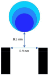
PNP calculations indicate that a sphere composed of 10 elemental positive charges whose lower edge is located 0.5 nm from the opening of a 0.9 nm diameter pore (5 nm in length) will produce a rectification of approximately 100 nS/100 mV in an aqueous medium containing 1 M monovalent salt. The spheres are 0.3, 0.4, and 0.5 nm in radius, each of which could produce the observed rectification. To save space, only a portion of the pore is illustrated.
Figure 14
Figure 14
Revised model of the gating of Triplin based on the results presented. The top of the structure is the cis side of the membrane, the side from which Triplin inserted. The bottom of the structure is the trans side and that is the side maintained at virtual ground by the amplifier. All indicated voltages refer to the cis side. The numbers 1,2,3 refer to pores 1,2 and 3. Short arrows refer to reactions of lower probability. For simplicity, the closed state of the pore is illustrated as the result of blockage by a single loop of the beta barrel but, of course, multiple loops may be involved.
Figure 15
Figure 15
Subunit 3 oscillations between the open and closed states terminate when subunit 2 opens. The left part of the figure shows the oscillations of subunit 3 held at 34 mV. At point (A), subunit 2 opens, thus, locking subunit 3 in its open state. At point (B), a triangular voltage wave was applied. Subunit 2 closure took place in the region labeled (C) and subunit 3 closure in the region labeled (D).

References

    1. Nikaido H. Molecular basis of bacterial outer membrane permeability revisited. Microbiol. Mol. Biol. Rev. 2003;67:593–656. doi: 10.1128/MMBR.67.4.593-656.2003. - DOI - PMC - PubMed
    1. Koebnik R., Locher K.P., Van Gelder P. Structure and function of bacterial outer membrane proteins: Barrels in a nutshell. Mol. Microbiol. 2000;37:239–253. doi: 10.1046/j.1365-2958.2000.01983.x. - DOI - PubMed
    1. Schindler H., Rosenbusch J.P. Matrix protein in planar membranes: Clusters of channels in a native environment and their functional reassembly. Proc. Natl. Acad. Sci. USA. 1981;78:2302–2306. doi: 10.1073/pnas.78.4.2302. - DOI - PMC - PubMed
    1. Van Gelder P., Saint N., Phale P., Eppens E.F., Prilipov A., van Boxtel R., Rosenbusch J.P., Tommassen J. Voltage sensing in the PhoE and OmpF outer membrane porins of Escherichia coli: Role of charged residues. J. Mol. Biol. 1997;269:468–472. doi: 10.1006/jmbi.1997.1063. - DOI - PubMed
    1. Robertson K.M., Tieleman D.P. Molecular basis of voltage gating of OmpF porin. Biochem. Cell Biol. 2002;80:517–523. doi: 10.1139/o02-145. - DOI - PubMed

LinkOut - more resources