. 2022 Nov 9;23(22):13797.
doi: 10.3390/ijms232213797.

PAC1, VPAC1, and VPAC2 Receptor Expression in Rat and Human Trigeminal Ganglia: Characterization of PACAP-Responsive Receptor Antibodies

Affiliations

PAC1, VPAC1, and VPAC2 Receptor Expression in Rat and Human Trigeminal Ganglia: Characterization of PACAP-Responsive Receptor Antibodies

Zoe Tasma et al. Int J Mol Sci. .

Abstract

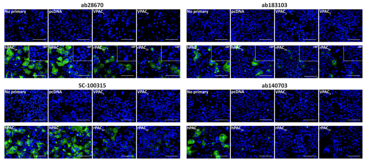
Pituitary adenylate cyclase-activating peptide (PACAP) is a neuropeptide expressed in the trigeminal ganglia (TG). The TG conducts nociceptive signals in the head and may play roles in migraine. PACAP infusion provokes headaches in healthy individuals and migraine-like attacks in patients; however, it is not clear whether targeting this system could be therapeutically efficacious. To effectively target the PACAP system, an understanding of PACAP receptor distribution is required. Therefore, this study aimed to characterize commercially available antibodies and use these to detect PACAP-responsive receptors in the TG. Antibodies were initially validated in receptor transfected cell models and then used to explore receptor expression in rat and human TG. Antibodies were identified that could detect PACAP-responsive receptors, including the first antibody to differentiate between the PAC1n and PAC1s receptor splice variants. PAC1, VPAC1, and VPAC2 receptor-like immunoreactivity were observed in subpopulations of both neuronal and glial-like cells in the TG. In this study, PAC1, VPAC1, and VPAC2 receptors were detected in the TG, suggesting they are all potential targets to treat migraine. These antibodies may be useful tools to help elucidate PACAP-responsive receptor expression in tissues. However, most antibodies exhibited limitations, requiring the use of multiple methodologies and the careful inclusion of controls.

Keywords: PAC1 receptor; PACAP; VIP; VPAC1 receptor; VPAC2 receptor; migraine; trigeminal ganglia.


Conflict of interest statement

Debbie L. Hay is or has been a consultant or speaker for Lilly, Amgen, Teva, Intarcia, Merck Sharp & Dohme and has received research funding from Living Cell Technologies (Sydney, Australia) and Abbvie Inc. (Mettawa, IL USA.) in the past three years. Christopher S. Walker has received research support from Living Cell Technologies and Abbvie Inc.

Figures

Figure 1
Figure 1
PAC1 receptor-like immunoreactivity detected in transfected Cos7 cells. Cells were transfected with the hPAC1n, hPAC1s, rPAC1n, rPAC1s, hVPAC1 and hVPAC2 receptors or vector (pcDNA3.1+). Cells were stained with ab28670 (1:1000) or subjected to antigen retrieval before addition of ab183103 (1:100), SC-100315 (1:50) or ab140703 (1:100). Pre-absorbed blocking peptide (BP) antibody controls for certain antibodies are presented as insets. Antibody immunoreactivity is shown in green and nuclear DAPI staining in blue. Images were taken with a 40× non-confocal objective and represent one field of view from three independent experiments. Scale bar, 100 µm.
Figure 2
Figure 2
Apparent PAC1 receptor molecular weights detected by anti-PAC1 receptor antibodies in transfected Cos7 cell protein. Protein samples were prepared from Cos7 cells transfected with the hPAC1n, hPAC1s, rPAC1n, rPAC1s, hVPAC1 and hVPAC2 receptors or vector (pcDNA3.1+). Protein was loaded, and blots probed with anti-PAC1 receptor antibody (1:1000). The exposure times were 7 s (1 µg) and 2 s (10 µg) for ab28670, 2.5 min (1 µg) and 31 s (10 µg) for ab183103, 15 s (1 µg) and 4 s (10 µg) for SC-100315, 28 s (1 µg) and 14 s (10 µg) for ab140703. Blots are representative of three (1 µg) or two (10 µg) independent experiments. Green arrowheads represent bands of an apparent MW consistent with the anti-HA PAC1 control (Figure S1C). Rat PAC1 receptor MWs were not clearly observed when 1 µg protein was loaded and are indicated on the 10 µg blot instead. A second green arrowhead indicates a receptor dimer. Pink arrowheads indicate major non-specific bands observed with 10 µg protein.
Figure 3
Figure 3
PAC1 receptor-like immunoreactivity detected in rat and human trigeminal ganglia. PAC1 (ab28670, 1:500; ab183103, ab140703, 1:100) receptor-like immunoreactivity in TG neurons and glia colocalized with β-tubulin (1:500). Pre-absorbed blocking peptide (BP) antibody controls are presented as insets. PAC1 antibody immunoreactivity is shown in pink, β-tubulin in green, colocalized regions appear white, and DAPI (rat) or Hoechst (human) stained nuclei in blue. Solid arrowheads indicate examples of immunoreactive neurons and empty arrowheads examples of immunoreactive glia. Images were taken with a 20× non-confocal objective and are representative of TG staining in three individual rat or human cases. Scale bar, 100 µm.
Figure 4
Figure 4
PAC1 receptor apparent molecular weights detected in rat and human trigeminal ganglia. Protein samples were prepared from TG or Cos7 cells transfected with the human or rat PAC1n and PAC1s receptors or vector (pcDNA3.1+). Protein was loaded, and blots probed with anti-PAC1 receptor antibodies (1:1000). The exposure times for ab28670 were 10 s (rat) and 7 s (human), ab183103 were 30 min (rat) and 16 min (human), SC-100315 were 16 s (rat) and 6 s (human), and ab140703 was 6 min. Blots are representative of TG protein prepared from three different rats or two human cases.
Figure 5
Figure 5
VPAC1 and VPAC2 receptor-like immunoreactivity detected in transfected Cos7 cells. Cells were transfected with the hPAC1n, hVPAC1 and hVPAC2 receptors or vector (pcDNA3.1+). Cells were stained with SC-377152 (1:200), SAB5500193, SAB5500194 (1:100, 1:200) or ab28624 (1:500, 1:1000). VPAC1 and VPAC2 receptor images shown were incubated with 1:100 SAB5500193 or SAB5500194, respectively and all other conditions with 1:200 antibody. The VPAC2 receptor image shown for ab28624 was incubated with 1:1000 antibody, and all other conditions with 1:500. Pre-absorbed blocking peptide (BP) antibody controls are presented as insets. Antibody immunoreactivity is shown in green and nuclear DAPI staining in blue. Images were taken with a 40× non-confocal objective and represent one field of view from three independent experiments. Scale bar, 100 µm.
Figure 6
Figure 6
Apparent VPAC1 and VPAC2 receptor molecular weights detected by anti-VPAC receptor antibodies in transfected Cos7 cell protein. Protein samples were prepared from Cos7 cells transfected with the hPAC1n, hVPAC1 and hVPAC2 receptors or vector (pcDNA3.1+). Protein was loaded, and blots probed with anti-VPAC1 or VPAC2 receptor antibody (1:1000). The exposure times were 5 s (1 µg) and 3 s (10 µg) for SC-377152, 7 s (1 µg) and 3 s (10 µg) for SAB5500193, 2 s (1 µg) and 3 s (10 µg) for ab28624 and 15 s (1 µg) and 4 s (10 µg) for SAB5500194. Blots are representative of three (1 µg) or two (10 µg) independent experiments. Green arrowheads represent bands of an apparent MW consistent with those observed in a prior study using protein from transfected HEK293 cells [35]. A second green arrowhead indicates a receptor dimer. Pink arrowheads indicate major non-specific bands observed with 10 µg protein.
Figure 7
Figure 7
VPAC1 receptor-like immunoreactivity detected in rat and human trigeminal ganglia. VPAC1 (SC-377152, SAB5500193, 1:100) receptor-like immunoreactivity in TG neurons and glia colocalized with β-tubulin (1:500). Pre-absorbed blocking peptide (BP) antibody controls are presented as insets. Receptor immunoreactivity is shown in pink, β-tubulin in green, colocalized regions appear white, and DAPI (rat) or Hoechst (human) stained nuclei in blue. Solid arrowheads indicate examples of immunoreactive neurons and empty arrowheads examples of immunoreactive glia. Images were taken with a 20× non-confocal objective and are representative of TG staining in three individual rat or human cases. Scale bar, 100 µm. * autofluorescence due to lipofuscin.
Figure 8
Figure 8
VPAC2 receptor-like immunoreactivity detected in rat and human trigeminal ganglia. VPAC2 (ab28624, 1:500, SAB5500194, 1:100) receptor-like immunoreactivity in TG neurons and glia colocalized with β-tubulin (1:500). Pre-absorbed blocking peptide (BP) antibody controls are presented as insets. Receptor immunoreactivity is shown in pink, β-tubulin in green, colocalized regions appear white, and DAPI (rat) or Hoechst (human) stained nuclei in blue. Solid arrowheads indicate examples of immunoreactive neurons and empty arrowheads examples of immunoreactive glia. Images were taken with a 20× non-confocal objective and are representative of TG staining in three individual rat or human cases. Scale bar, 100 µm. * autofluorescence due to lipofuscin.
Figure 9
Figure 9
VPAC1 and VPAC2 receptor apparent molecular weights detected in rat and human trigeminal ganglia. Protein samples were prepared from TG or Cos7 cells transfected with the human PAC1n, VPAC1 and VPAC2 receptors or vector (pcDNA3.1+). Protein was loaded, and blots probed with anti-VPAC receptor antibodies (1:1000). The exposure times for SC-377152 was 16 s, SAB5500193 were 8 min (rat) and 3 min (human), ab28624 was 14 s, and SAB5500194 were 15 s (rat) and 8 s (human). Blots are representative of TG protein prepared from three different rats or two human cases.

Similar articles

Cited by

References

    1. Dun N., Miyazaki T., Tang H., Dun E. Pituitary adenylate cyclase activating polypeptide immunoreactivity in the rat spinal cord and medulla: Implication of sensory and autonomic functions. Neuroscience. 1996;73:677–686. doi: 10.1016/0306-4522(96)00057-7. - DOI - PubMed
    1. Jansen-Olesen I., Baun M., Amrutkar D.V., Ramachandran R., Christophersen D.V., Olesen J. PACAP-38 but not VIP induces release of CGRP from trigeminal nucleus caudalis via a receptor distinct from the PAC1 receptor. Neuropeptides. 2014;48:53–64. doi: 10.1016/j.npep.2014.01.004. - DOI - PubMed
    1. Kummer W., Heym C. Correlation of neuronal size and peptide immunoreactivity in the guinea-pig trigeminal ganglion. Cell Tissue Res. 1986;245:657–665. doi: 10.1007/BF00218569. - DOI - PubMed
    1. Uddman R., Tajti J., Hou M., Sundler F., Edvinsson L. Neuropeptide Expression in the Human Trigeminal Nucleus Caudalis and in the Cervical Spinal Cord C1 and C2. Cephalalgia. 2002;22:112–116. doi: 10.1046/j.1468-2982.2002.00324.x. - DOI - PubMed
    1. Csati A., Tajti J., Kuris A., Tuka B., Edvinsson L., Warfvinge K. Distribution of vasoactive intestinal peptide, pituitary adenylate cyclase-activating peptide, nitric oxide synthase, and their receptors in human and rat sphenopalatine ganglion. Neuroscience. 2012;202:158–168. doi: 10.1016/j.neuroscience.2011.10.055. - DOI - PubMed

MeSH terms

Substances