. 2022 Nov 10;23(22):13849.
doi: 10.3390/ijms232213849.

Inhibition of Calpain Attenuates Degeneration of Substantia Nigra Neurons in the Rotenone Rat Model of Parkinson's Disease

Affiliations

Inhibition of Calpain Attenuates Degeneration of Substantia Nigra Neurons in the Rotenone Rat Model of Parkinson's Disease

Vandana Zaman et al. Int J Mol Sci. .

Abstract

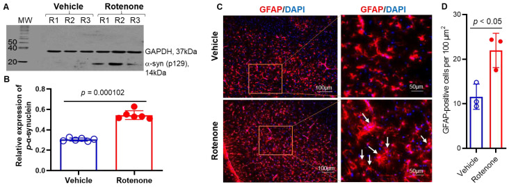
In the central nervous system (CNS), calcium homeostasis is a critical determinant of neuronal survival. Calpain, a calcium-dependent neutral protease, is widely expressed in the brain, including substantia nigra (SN) dopaminergic (DA) neurons. Though calpain is implicated in human Parkinson's disease (PD) and corresponding animal models, the roles of specific ubiquitous calpain isoforms in PD, calpain-1 and calpain-2, remain poorly understood. In this study, we found that both isoforms are activated in a nigrostriatal pathway with increased phosphorylated synuclein following the administration of rotenone in Lewis rats, but calpain isoforms played different roles in neuronal survival. Although increased expression of calpain-1 and calpain-2 were detected in the SN of rotenone-administered rats, calpain-1 expression was not altered significantly after treatment with calpain inhibitor (calpeptin); this correlated with neuronal survival. By contrast, increased calpain-2 expression in the SN of rotenone rats correlated with neuronal death, and calpeptin treatment significantly attenuated calpain-2 and neuronal death. Calpain inhibition by calpeptin prevented glial (astroglia/microglia) activation in rotenone-treated rats in vivo, promoted M2-type microglia, and protected neurons. These data suggest that enhanced expression of calpain-1 and calpain-2 in PD models differentially affects glial activation and neuronal survival; thus, the attenuation of calpain-2 may be important in reducing SN neuronal loss in PD.

Keywords: Parkinson’s disease; alpha-synuclein; calpain; dopaminergic neuron; microglia; rotenone; substantia nigra.


Conflict of interest statement

The authors have no financial conflict of interest.

Figures

Figure 1
Figure 1
Administration of rotenone-induced increased expression of p-α syn 129 in the dorsal striatum of Lewis rats. (A) A representative photomicrograph of Western blot analysis showed increased expression of p-α-syn 129 in the striatum. GAPDH was used as a loading control. MW = molecular weight in kDa. (B) Densitometric analysis of p-α-syn 129 protein expression by ImageJ software shows that there is a significant increase (p < 0.001) in the synuclein protein expression in rotenone rats. N = 6. (C) Presence of highly active GFAP (red)-immunostained astrocytes in the SN following rotenone administration in rats. DAPI (blue) was used for nuclear staining. Representative microphotographs from control and rotenone groups suggest GFAP-immunostained astrocytes are hypertrophied in appearance in rotenone-injected rats as compared to vehicle controls. (D) Quantitative analysis of GFAP+ cells by cellSens Imaging Software suggests that astrocyte numbers were significantly increased following rotenone injection. N = 3.
Figure 2
Figure 2
Rotenone-induced distinct loss of SN DA neurons and fibers. (A) Immunohistochemistry of rat SN samples with TH (red) following rotenone administration and calpain inhibition by CP treatment. Distinct loss of TH-positive neurons and fibers were seen in the rotenone rats as compared to controls. DAPI (blue) was used for nuclear staining. Note the dense TH staining of neurons in the SNpc (arrows) and DA fibers in SNpr (arrowheads) in the control group and also in rotenone plus CP treatment group. The extensive loss of TH-labeled neurons (arrows) and fibers (arrowheads) was demonstrated after rotenone administration. However, calpeptin treatment (rotenone+ CP) prevented neuronal loss in SN. (B) Analysis of TH-immunostained fiber density in SNpr by ImageJ software shows that there was a significant decrease (* p < 0.001) in TH fiber density in rotenone rats, which was prevented by CP treatment. N = 3.
Figure 3
Figure 3
Calpain-1 expression was upregulated in SN DA neurons following rotenone administration, and it was retained after CP treatment. (A) Representative images from the respective treatment groups showed that TH-immunostained SN DA neurons co-localized with calpain-1 (arrows). Calpain-1 staining (red) indicates cytoplasmic localization in the SN neurons stained with TH (green). Merged images showed co-localization (yellow) of TH+ calpain-1 with a distinctly DAPI counter-stained nucleus (blue/purple color) after CP treatment. N = 3. (B) Quantitative analysis of TH+ and calpain-1+ cells colocalized per 100 µm2 following CP treatment. N = 4–5.
Figure 4
Figure 4
Calpain-2 expression was upregulated in SN DA neurons following rotenone administration, and it was not retained after CP treatment. (A) Representative images from the respective treatment groups showed SN DA neurons immunostained with TH (green) and calpain-2 (red). Co-localization of TH and calpain-2 (merged images) with DAPI (blue/purple) as a counter stain. Merged images showed less co-localization (yellow) of TH+ calpain-2 with a distinctly DAPI counter-stained nucleus (blue/purple color) after CP treatment. N = 3. (B) Quantitative analysis of TH+ and calpain-2+ cells colocalized per 100 µm2 following CP treatment. N = 4.
Figure 5
Figure 5
Activated astrocytes in the rat dorsal striatum following rotenone administration. Representative photomicrographs demonstrate GFAP-labeled astrocytes in the striatum in control, rotenone, and rotenone + CP treatment groups. (A) Rotenone administration conspicuously increased the number and size of the astrocytes in the striatum, and treatment of rats with CP decreased the number of reactive astrocytes. (B) Quantitative analysis by cellSens Imaging Software showed that the number of reactive astrocytes was significantly increased (p = 0.0131) in rotenone vs. controls, whereas it was markedly decreased (p = 0.0314) following CP treatment. N = 3. (C,D) Immunohistochemistry with Iba1 showed that the microglial population was significantly increased (p = 0.0105) in the SN after rotenone administration of rats, which were not significantly changed (p = 0.0535) following CP treatment. (E) Differentiation of microglia in rotenone rats after CP treatment. Colocalization (yellow, arrows) of Iba1 (red) and arginase 1 (green) in the dorsal striatum of CP-treated rats indicated differentiation of microglia into M2-type following calpain inhibition. N = 3–5. (F) Quantitative analyses of co-localized Iba-1+- and Arginase 1+-stained cells from Figure 5E. N = 4.
Figure 6
Figure 6
Administration of rotenone differentially influenced the expression levels of calpain-1 and calpain-2 in the nigrostriatal pathway in rats. (A) A representative photomicrograph of Western blot analysis showed upregulation of calpain-1 protein expression in the striatum following rotenone injection in rats (upper panel). β-actin was used as a loading control. Quantification of the protein level detected in Western blot analysis suggests calpain-1 expression was significantly increased (p = 0.0005) after rotenone injection, and it was marginally decreased (p = 0.1171) following CP treatment (lower panel, N = 5–6). (B) A representative photomicrograph of Western blot analysis also showed upregulation of calpain-2 protein expression in the striatum following rotenone injection (upper panel). β-actin was used as a loading control. Quantification of the calpain-2 protein level suggests a significant increase (p < 0.0001) in calpain-2 protein expression in the rotenone rats as compared to control group (lower panel, N = 6–8). CP treatment significantly decreased calpain-2 protein expression (p < 0.0001) in rotenone rats. MW = molecular weight.
Figure 7
Figure 7
Schematic presentation showing rotenone-induced toxicity in SN neurons. Reactive astrocytes/microglia were detected after rotenone administration in Lewis rats. Both calpain-1 and calpain-2 were also activated in the nigrostriatal pathway following rotenone administration in rats. Calpain inhibition may help reduce glial activation via reduction of ROS and differentiation of microglia into M2-type cells. Decreased phagocytic function of glia and selective autophagy may also support preferential regulation of calpain-1 and calpain-2 expression/activity and the fate of neuronal survival in PD.

References

    1. Wong Y.C., Krainc D. alpha-synuclein toxicity in neurodegeneration: Mechanism and therapeutic strategies. Nat. Med. 2017;23:1–13. doi: 10.1038/nm.4269. - DOI - PMC - PubMed
    1. Zaman V., Shields D.C., Shams R., Drasites K.P., Matzelle D., Haque A., Banik N.L. Cellular and molecular pathophysiology in the progression of Parkinson’s disease. Metab. Brain Dis. 2021;36:815–827. - PMC - PubMed
    1. Liang C.L., Sinton C.M., German D.C. Midbrain dopaminergic neurons in the mouse: Co-localization with Calbindin-D28K and calretinin. Neuroscience. 1996;75:523–533. doi: 10.1016/0306-4522(96)00228-X. - DOI - PubMed
    1. Nemoto C., Hida T., Arai R. Calretinin and calbindin-D28k in dopaminergic neurons of the rat midbrain: A triple-labeling immunohistochemical study. Brain Res. 1999;846:129–136. doi: 10.1016/S0006-8993(99)01950-2. - DOI - PubMed
    1. German D.C., Manaye K.F., Sonsalla P.K., Brooks B.A. Midbrain dopaminergic cell loss in Parkinson’s disease and MPTP-induced parkinsonism: Sparing of calbindin-D28k-containing cells. Ann. N. Y. Acad. Sci. 1992;648:42–62. doi: 10.1111/j.1749-6632.1992.tb24523.x. - DOI - PubMed