Review
. 2022 Nov 10;23(22):13850.
doi: 10.3390/ijms232213850.

The Role of Copper Homeostasis in Brain Disease

Affiliations
Review

The Role of Copper Homeostasis in Brain Disease

Yumei An et al. Int J Mol Sci. .

Abstract

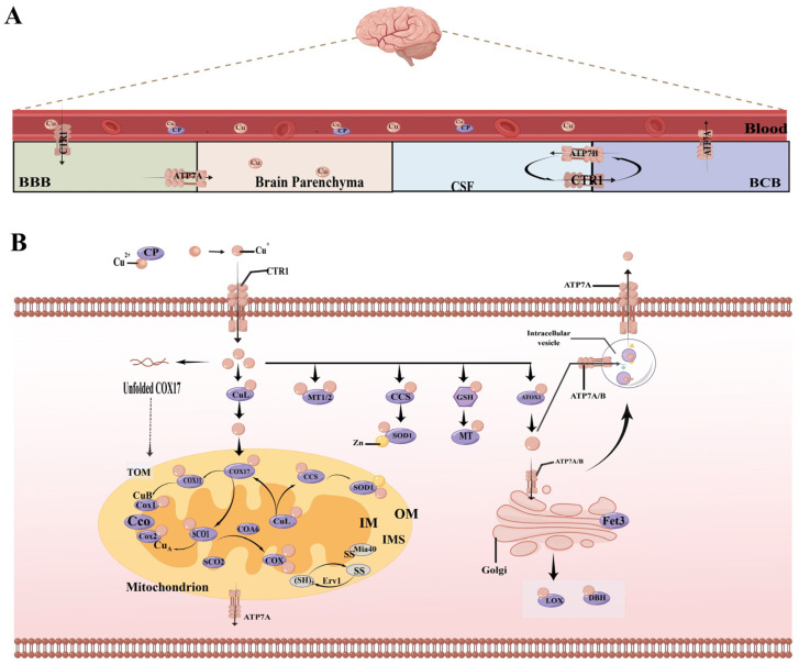
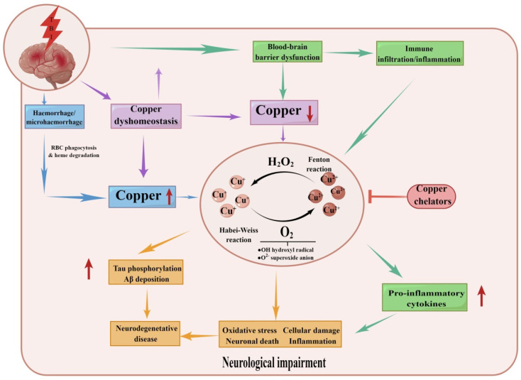
In the human body, copper is an important trace element and is a cofactor for several important enzymes involved in energy production, iron metabolism, neuropeptide activation, connective tissue synthesis, and neurotransmitter synthesis. Copper is also necessary for cellular processes, such as the regulation of intracellular signal transduction, catecholamine balance, myelination of neurons, and efficient synaptic transmission in the central nervous system. Copper is naturally present in some foods and is available as a dietary supplement. Only small amounts of copper are typically stored in the body and a large amount of copper is excreted through bile and urine. Given the critical role of copper in a breadth of cellular processes, local concentrations of copper and the cellular distribution of copper transporter proteins in the brain are important to maintain the steady state of the internal environment. The dysfunction of copper metabolism or regulatory pathways results in an imbalance in copper homeostasis in the brain, which can lead to a myriad of acute and chronic pathological effects on neurological function. It suggests a unique mechanism linking copper homeostasis and neuronal activation within the central nervous system. This article explores the relationship between impaired copper homeostasis and neuropathophysiological progress in brain diseases.

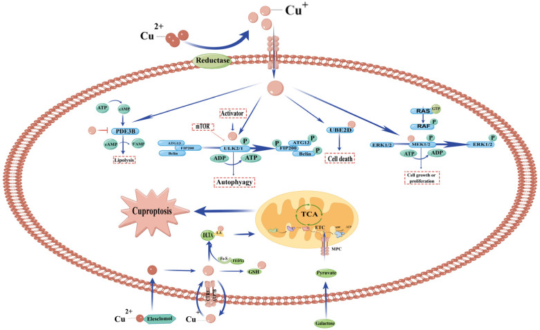
Keywords: brain injury; cognition; copper; cuproptosis; neurodegeneration.


Conflict of interest statement

The authors declare no conflict of interest.

Figures

Figure 1
Figure 1
(A) The entry and exit of copper in the brain. Copper enters the brain through the blood–brain barrier (BBB). The endothelial cells that make up the BBB get copper from the blood via the apical copper transporter1 (CTR1) and transport it to the brain parenchyma via ATP7A. If there is an excess of copper, then the excess copper is released from the brain cells into the cerebrospinal fluid (CSF) and is taken up by the cells that make up the blood–cerebrospinal fluid barrier (BCB). The copper taken up by these cells is either stored by ATP7B for potential transport to the CSF or transported into the blood by ATP7A. (B) Copper metabolism of brain cells. Ceruloplasmin (CP) carries the copper to its destination. On the plasma membrane, copper ion channel CTR1 can achieve a high affinity for copper uptake. After copper enters the cell, a small copper ligand (CuL) supplies Cu+ to the mitochondria intermembrane space (IMS). In the mitochondria, copper chaperone for cytochrome C oxidase 17 (COX17) supplies two pathways, delivering copper to COX11 and synthesis of cytochrome oxidase1 (SCO1). Copper reaches the CuB site of the COX1 subunit via COX11 and the CuA site of COX2 via SCO1, participating in the metallization of the mitochondrial cytochrome C oxidase (CCO) complex and embedded in the inner membrane (IM). Nuclear encoded mitochondrial proteins, unfolded COX17, are imported across the outer membrane (OM) unfolded via the TOM translocase and then captured in the inner membrane space (IMS), following the introduction of disulfide bonds (SS) through the actions of Mia40. A sulfhydryl oxidase Erv1 generates a reactive disulfide on Mia40. Copper chaperone for cytochrome c oxidase (COX) is catalyzed by SCO1 and SCO2 which are metallochaperones. Cytochrome c oxidase assembly factor 6 (COA6) and SCO2 assist in keeping the redox balance of SCO1, which in turn helps maintain its copper binding and transport to COX. In the cytoplasm, metallothionein 1/2 (MT1/2) binds to more than one copper ion and can act as a reservoir for copper. Copper chaperone for superoxide dismutase (CCS) delivers copper to Cu/Zn superoxide dismutase (SOD). In addition, glutathione (GSH) can also be directly or indirectly involved in regulating the cellular copper pool. Copper ions bind to antioxidant protein 1 (Atox1), which presents copper to the ATP-driven transmembrane copper ion pumps ATP7A and ATP7B, both of which perform both copper export and metallochaperone functions, with ATP7B performing copper export in hepatocytes and ATP7A primarily performing copper export in brain cells. Together these proteins maintain proper intracellular copper bioavailability and ensure the metalation of copper-dependent enzymes including COX, superoxide dismutase 1 (SOD1) and oxygenases/oxidases including tyrosinase, lysine oxidase (LOX), dopamine β-hydroxylase (DBH). The figures in this article are all drawn by Figdraw.
Figure 2
Figure 2
Copper participates in cell death and proliferation pathways. Copper binds to and inhibits Phosphodiesterase 3b (PDE3B), inhibits cyclic AMP (cAMP) degradation, and promotes cAMP-dependent lipolysis, which is needed for fat metabolism. Copper-dependent kinase signalling can regulate autophagy through ULK1 and ULK2. The copper signal promotes protein degradation by binding the E2-binding enzyme UBE2D1-UBE2D4. Copper-dependent kinase signalling can regulate cell growth/proliferation through MEK1 and MEK2. Furthermore copper binds directly to the lipoylated components of the TCA cycle. The accumulation of these copper-bound lipoacylated mitochondrial proteins and the following loss of Fe-S cluster proteins then triggered cuproptosis.
Figure 3
Figure 3
The outcomes of imbalanced copper balance after TBI. TBI leads to several serious consequences, including BBB breakdown, haemorrhage, and copper dyshomeostasis. Together this leads to a copper increase or decrease in the brain. Copper is involved in the Haber–Weiss/Fenton reaction, promoting oxidative stress, neuronal death, inflammation onset and tau phosphorylation/beta deposition. This leads to pathological changes in traumatic brain injury and ultimately increases the risk of neurological decline and neurodegenerative disease.

References

    1. Rihel J. Copper on the brain. Nat. Chem. Biol. 2018;14:638–639. doi: 10.1038/s41589-018-0089-1. - DOI - PubMed
    1. Burkhead J.L., Gogolin Reynolds K.A., Abdel-Ghany S.E., Cohu C.M., Pilon M. Copper homeostasis. New Phytol. 2009;182:799–816. doi: 10.1111/j.1469-8137.2009.02846.x. - DOI - PubMed
    1. Kim B.E., Turski M.L., Nose Y., Casad M., Rockman H.A., Thiele D.J. Cardiac copper deficiency activates a systemic signaling mechanism that communicates with the copper acquisition and storage organs. Cell Metab. 2010;11:353–363. doi: 10.1016/j.cmet.2010.04.003. - DOI - PMC - PubMed
    1. Kim B.E., Nevitt T., Thiele D.J. Mechanisms for copper acquisition, distribution and regulation. Nat. Chem. Biol. 2008;4:176–185. doi: 10.1038/nchembio.72. - DOI - PubMed
    1. Ge E.J., Bush A.I., Casini A., Cobine P.A., Cross J.R., DeNicola G.M., Dou Q.P., Franz K.J., Gohil V.M., Gupta S., et al. Connecting copper and cancer: From transition metal signalling to metalloplasia. Nat. Rev. Cancer. 2022;22:102–113. doi: 10.1038/s41568-021-00417-2. - DOI - PMC - PubMed