. 2022 Nov 12;23(22):13984.
doi: 10.3390/ijms232213984.

2cChIP-seq and 2cMeDIP-seq: The Carrier-Assisted Methods for Epigenomic Profiling of Small Cell Numbers or Single Cells

Affiliations

2cChIP-seq and 2cMeDIP-seq: The Carrier-Assisted Methods for Epigenomic Profiling of Small Cell Numbers or Single Cells

Congxia Hu et al. Int J Mol Sci. .

Abstract

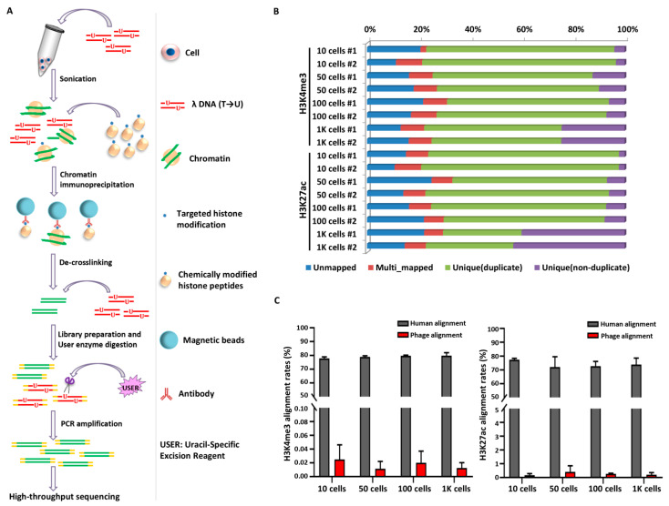
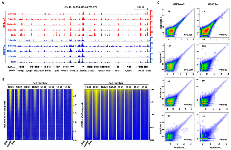
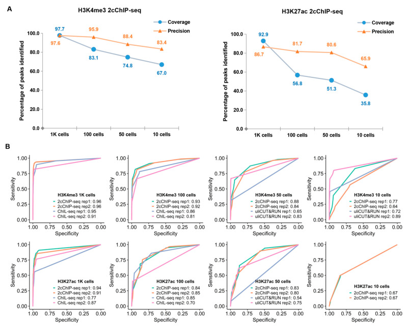
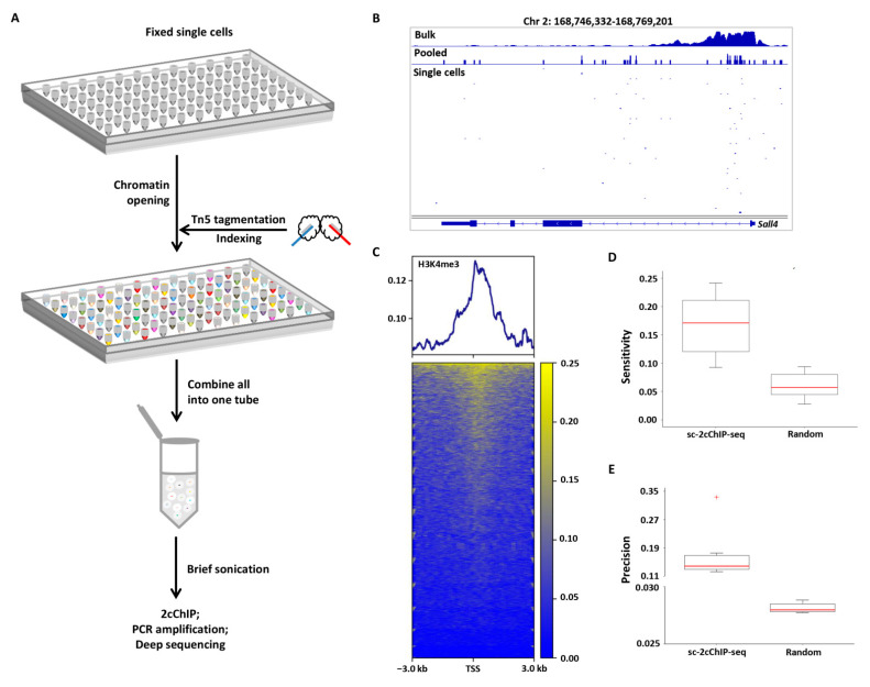
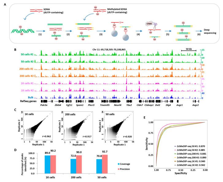
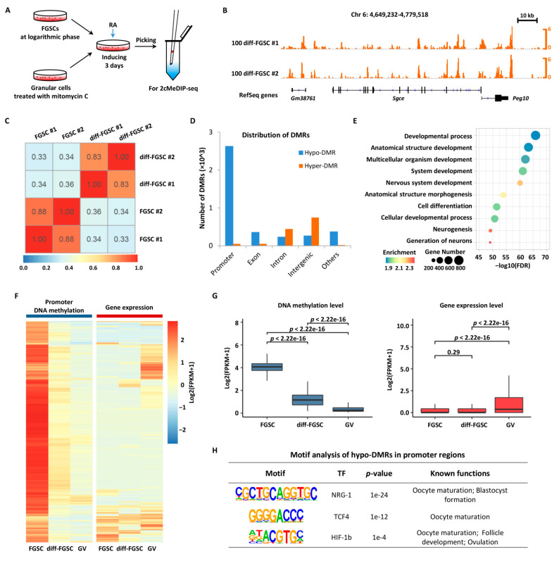
Chromatin immunoprecipitation coupled with high-throughput sequencing (ChIP-seq) can profile genome-wide epigenetic marks associated with regulatory genomic elements. However, conventional ChIP-seq is challenging when examining limited numbers of cells. Here, we developed a new technique by supplementing carrier materials of both chemically modified mimics with epigenetic marks and dUTP-containing DNA fragments during conventional ChIP procedures (hereafter referred to as 2cChIP-seq), thus dramatically improving immunoprecipitation efficiency and reducing DNA loss of low-input ChIP-seq samples. Using this strategy, we generated high-quality epigenomic profiles of histone modifications or DNA methylation in 10-1000 cells. By introducing Tn5 transposase-assisted fragmentation, 2cChIP-seq reliably captured genomic regions with histone modification at the single-cell level in about 100 cells. Moreover, we characterized the methylome of 100 differentiated female germline stem cells (FGSCs) and observed a particular DNA methylation signature potentially involved in the differentiation of mouse germline stem cells. Hence, we provided a reliable and robust epigenomic profiling approach for small cell numbers and single cells.

Keywords: female germline stem cells; low-input ChIP-seq; low-input MeDIP-seq; single-cell ChIP-seq.


Conflict of interest statement

The authors have declared no conflict of interest.

Figures

Figure 1
Figure 1
2cChIP-seq strategy overview and data quality presentation. (A) The scheme illustrates the conceptual design and key steps of the 2cChIP-seq protocol. The dUTP-containing lambda DNA fragments and chemically modified histone peptides are added as carriers during ChIP DNA preparation and library generation. (B) Deep sequencing features of 2cChIP-seq reads. (C) The plot depicts the proportions of 2cChIP-seq reads aligned to the human or lambda reference genome. Data were obtained from two independent experiments. The labels (#1 and #2) represent two 2cChIP-seq replicates.
Figure 2
Figure 2
2cChIP-seq reliably generates H3K4me3 and H3K27ac profiles in small numbers of cells. (A) Normalized H3K4me3 and H3K27ac 2cChIP-seq signals at the indicated regions using data from various sample sizes. ENCODE data (H3K4me3, GSM2534289; H3K27ac, and GSM733656) are shown for comparison. (B) Heatmaps of 2cChIP-seq data for the indicated cell numbers. Data are centered on peaks called from GSM2534289 for H3K4me3 and GSM733656 for H3K27ac. A window of 6 kb (−3 kb to +3 kb) around the peak center is shown. (C) Scatter plot comparison of two 2cChIP-seq biological replicate datasets generated with various numbers of cells. The r indicates Pearson’s correlation coefficient calculated in non-overlapped 4-kb bins across the entire human genome. Sample size: n = 2. Chr, Chromosome. #1 and #2, represent two 2cChIP-seq replicates.
Figure 3
Figure 3
Performance comparison between 2cChIP-seq and other epigenomic profiling methods. (A) Line charts showing the coverage and precision of 2cChIP-seq peaks in the promoter region compared with those of ENCODE ChIP-seq data (H3K4me3, GSM2534289; H3K27ac, GSM733656). (B) ROC curves for the comparison of H3K4me3 and H3K27ac, respectively. ROC curves were constructed by comparing the data generated by distinct methods for low sample input with published data. The raw data of ChIL-seq and uliCUT&RUN are from GSE115047 and GSE111121, respectively. The values shown are for the area under the ROC curve (AUC).
Figure 4
Figure 4
2cChIP-seq profiles of H3K4me3 signals in single cells. (A) Workflow of single-cell 2cChIP-seq. (B) Integrative Genomics Viewer (IGV) track view showing the H3K4me3 profiles from ENCODE bulk-cell ChIP-seq data (GSM1003756), pooled single-cell 2cChIP-seq data (from two independent experiments), and 12 individual cells. (C) Heatmap showing H3K4me3 signals at the TSS ±3 kb regions of genes for all single cells. (D,E) Boxplots showing the scores of sensitivity (D) and precision (E) for the top 5% of individual single cells, and simulated random genomic regions.
Figure 5
Figure 5
2cMeDIP-seq generates high-quality DNA methylone profiles with limited numbers of cells. (A) Overview of 2cMeDIP-seq protocol. Following (1) sample preparation and sonication, dUTP-containing λ DNA (lambda DNA) fragments are added as carrier, (2) repair of DNA ends and ligation of barcoded adaptors, (3) immunoprecipitation with antibody, methylated λ DNA (dUTP-containing) fragments are added as carrier, (4) reverse-crosslinking, (5) elution and USER digestion, (6) PCR amplification and deep sequencing. (B) Normalized 2cMeDIP-seq data generated with distinct numbers of FGSCs are shown for the indicated region. The ‘bulk’ data generated with millions of cells using the MeDIP-seq protocol is shown for comparison. (C) Scatter plot comparison of two 2cMeDIP-seq biological replicate datasets generated with various numbers of cells. The r indicates Pearson’s correlation coefficient calculated as in Figure 2C. (D) Bar chart showing coverage and precision of 2cMeDIP-seq signals in the indicated numbers of cells compared with the bulk counterpart. (E) ROC curves for the comparison for the 2cMeDIP-seq data generated by distinct numbers of cells. Values shown are AUC. Chr, chromosome. #1 and #2, two biological replicates.
Figure 6
Figure 6
Distinct DNA methylation patterns during FGSC differentiation. (A) Experimental design and DNA methylation data generated with differentiated FGSCs. FGSCs were treated with RA for 3 days and collected for 2cMeDIP-seq. (B) Normalized 2cMeDIP-seq signals generated with 100 differentiated FGSCs are shown for the indicated region. (C) Heatmap showing the methylation signals correlation between the undifferentiated and the differentiated FGSCs. The correlation coefficient was calculated in non-overlapping 10 kb windows across entire genome. (D) The bar chart showing the numbers of DMRs distributed in distinct genomic regions of differentiated FGSCs when compared with the undifferentiated FGSCs [FDR (false discovery rate) < 0.001]. (E) GO analysis of genes with hypo-DMRs in promoter regions. The color and the dot size represent the enrichment levels and the number of DMR-associated genes within each GO term, respectively. (F) DNA methylation and expression analysis of genes with hypomethylated promoters. (G) Boxplots of DNA methylation or gene expression levels of the genes with hypomethylated promoters in FGSCs, diff-FGSCs, and GV oocytes. (H) Motifs analysis of hypomethylated DMRs in promoter regions.

Similar articles

Cited by

References

    1. Kharchenko P.V., Tolstorukov M.Y., Park P.J. Design and analysis of ChIP-seq experiments for DNA-binding proteins. Nat. Biotechnol. 2008;26:1351–1359. doi: 10.1038/nbt.1508. - DOI - PMC - PubMed
    1. Rodríguez-Ubreva J., Ballestar E. Chromatin immunoprecipitation. Methods Mol. Biol. 2014;1094:309–318. doi: 10.1007/978-1-62703-706-8_24. - DOI - PubMed
    1. Brind’Amour J., Liu S., Hudson M., Chen C., Karimi M.M., Lorincz M.C. An ultra-low-input native ChIP-seq protocol for genome-wide profiling of rare cell populations. Nat. Commun. 2015;6:6033. doi: 10.1038/ncomms7033. - DOI - PubMed
    1. Gilfillan G.D., Hughes T., Sheng Y., Hjorthaug H.S., Straub T., Gervin K., Harris J.R., Undlien D.E., Lyle R. Limitations and possibilities of low cell number ChIP-seq. BMC Genom. 2012;13:645. doi: 10.1186/1471-2164-13-645. - DOI - PMC - PubMed
    1. Dahl J.A., Collas P. Q2ChIP, a quick and quantitative chromatin immunoprecipitation assay, unravels epigenetic dynamics of developmentally regulated genes in human carcinoma cells. Stem Cells. 2007;25:1037–1046. doi: 10.1634/stemcells.2006-0430. - DOI - PubMed

LinkOut - more resources