. 2022 Nov 13;23(22):14008.
doi: 10.3390/ijms232214008.

(-)-Epigallocatechin-3-Gallate Prevents IL-1β-Induced uPAR Expression and Invasiveness via the Suppression of NF-κB and AP-1 in Human Bladder Cancer Cells

Affiliations

(-)-Epigallocatechin-3-Gallate Prevents IL-1β-Induced uPAR Expression and Invasiveness via the Suppression of NF-κB and AP-1 in Human Bladder Cancer Cells

Dhiraj Kumar Sah et al. Int J Mol Sci. .

Abstract

(-)-Epigallocatechin-3-O-gallate (EGCG), a primary green tea polyphenol, has powerful iron scavengers, belongs to the family of flavonoids with antioxidant properties, and can be used to prevent cancer. Urokinase-type plasminogen activator receptors (uPARs) are glycosylphosphatidylinositol (GPI)-anchored cell membrane receptors that have crucial roles in cell invasion and metastasis of several cancers including bladder cancer. The mechanism of action of EGCG on uPAR expression has not been reported clearly yet. In this study, we investigated the effect of EGCG on interleukin (IL)-1β-induced cell invasion and uPAR activity in T24 human bladder cancer cells. Interestingly, nuclear factor (NF)-κB and activator protein (AP)-1 transcription factors were critically required for IL-1β-induced high uPAR expression, and EGCG suppressed the transcriptional activity of both the ERK1/2 and JNK signaling pathways with the AP-1 subunit c-Jun. EGCG blocked the IL-1β-stimulated reactive oxygen species (ROS) production, in turn suppressing NF-κB signaling and anti-invasion effects by inhibiting uPAR expression. These results suggest that EGCG may exert at least part of its anticancer effect by controlling uPAR expression through the suppression of ERK1/2, JNK, AP-1, and NF-κB.

Keywords: AP-1; EGCG; IL-1β; NF-κB; ROS; Urokinase-type plasminogen activator receptor (uPAR); cell invasion.


Conflict of interest statement

The authors declare no conflict of interest.

Figures

Figure 1
Figure 1
(A) Expression of uPAR (i) and IL1B (ii) in TPM scores of bladder cancer data obtained from GEPIA 2. (B) Expression of uPAR in non-papillary and papillary bladder cancer (p < 0.05). (C) uPAR expression in various stages of bladder cancer. Expression correlation and protein interaction between IL1B, NFKB1, JUN, and MAPK1 with PLAUR in BLCA data obtained from TIMER2.0 (p < 0.05) (D) and GeneMANIA (E).
Figure 2
Figure 2
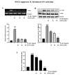
EGCG inhibits IL-1β-induced uPAR expression in T24 bladder cancer cells. (A) T24 cells were treated with 0−10 ng/mL of IL-1β for 4 h, and uPAR mRNA level was evaluated by RT-PCR. The bladder cancer cell line T24 was pretreated with different concentration of EGCG (5–50 μM) for 1 h. The cells were then incubated with IL-1β (5 ng/ ml) for 4 h. Then, mRNA was extracted and uPAR expression was evaluated by RT-PCR (B). (C,D) T24 cells were treated with 0–10 ng/mL IL-1β for 4 h, and uPAR protein level was evaluated by Western blotting. The T24 cell line was pretreated with different concentration of EGCG (5–50 μM) for 1 h. The cells were then incubated with IL-1β (5 ng/ ml) for 4 h. Then, protein was extracted and uPAR expression was evaluated by Western blotting. (E) T24 cells transfected with the pGL3-uPAR plasmid were pretreated with 5–50 μM for 1 h then incubated with 5 ng/ mL IL-1β for 12 h submitted for the luciferase assay. The above data represent the mean ± SD from triplicate measurements (# p < 0.05, #### p < 0.0001 versus control; * p < 0.05, ** p < 0.01, *** p < 0.001, **** p < 0.0001 versus IL-1β).
Figure 3
Figure 3
EGCG inhibits MAPK signaling stimulated by IL-1β in T24 bladder cancer cells (A) RT-PCR analysis to detect the uPAR expression by 1 h pretreatment with signaling inhibitor and then 5 ng/mL IL-1β treatment for 4 h. (B) T24 cells pretreated with 10–50 μM EGCG for 1 h were exposed to 5 ng/mL IL-1β for 30 min. The cells were then extracted for protein and tested for the level of phosphorylated ERK1/2, JNK and P38. (C) Phosphorylation of c-Jun by pre-treatment with MAPK inhibitors for 1 h were exposed to 5 ng/mL IL-1β for 30 min. The above data represent the mean ± SD from triplicate measurements (# p < 0.05, ## p < 0.01, #### p < 0.0001 versus control; ** p < 0.01, *** p < 0.001 versus IL-1β).
Figure 4
Figure 4
EGCG inhibits IL-1β-induced uPAR expression by suppressing the transcriptional activity of AP-1 in T24 cells. (A) T24 cells were pretreated with EGCG (10, 30, and 50 μM) for 1 h and then 5 ng/mL IL-1β for 4 h, and the cells were then extracted for mRNA to check uPAR expression by RT-PCR for the c-Jun expression. T24 cells were pretreated again with different concentrations of EGCG followed by 5 ng/mL IL-1β for 30 min of treatment and the phosphorylation of c-Jun expression by performing Western blotting (B). (C) Cells were pretreated with EGCG (10, 30, and 50 μM), were transiently transfected with the AP-1 luciferase reporter plasmid and were incubated with 5 ng/mL IL-1β; the cells were lysed, and luciferase activity was determined. The above data represent the mean ± SD from triplicate measurements (### p < 0.001, #### p < 0.0001 versus control; * p < 0.05, ** p < 0.01, *** p < 0.001, **** p < 0.0001 versus IL-1β).
Figure 5
Figure 5
EGCG Suppresses IL-1β-induced NF-κB signaling in T24 cells. (A) T24 cells were pre-treated with 10–50 µM EGCG and incubated with 5 ng/mL IL-1β for 4 h; cells were then harvested to check for p65 and IκBα by Western blot analysis. (B) Cells were pretreated with EGCG (10, 30, and 50 μM), were transiently transfected with the AP-1 luciferase reporter plasmid and were incubated with 5 ng/mL IL-1β; the cells were lysed, and luciferase activity was determined. (C) T24 cells were pretreated with 10–20 µM Bay and incubated with 5 ng/mL IL-1β for 4 h. The cells were then extracted for mRNA, and uPAR expression was checked by RT-PCR. (D) T24 cells were pretreated with 5–20 mM NAC for 1 h and incubated with 5 ng/mL IL-1β for 4 h, cells were then harvested to check for p-NF-κB and p-IκBα by Western blot analysis. The above data represent the mean ± SD from triplicate measurements (# p < 0.05, ## p < 0.01, #### p < 0.0001 versus control; * p < 0.05, ** p < 0.01, *** p < 0.001, **** p < 0.0001 versus IL-1β).
Figure 6
Figure 6
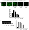
EGCG blocks ROS production induced by IL-1β in T24 cells. (A) Representative images (200X) and statistically quantitative values of ROS production by confocal microscope. (B) Relative fluorescence intensity fold by EGCG and IL-1β. (C) RT-PCR analysis for uPAR expression on NAC (5–20 mM) pretreatment for 1 h followed by 5 ng/mL IL-1β treatment for 4 h. The above data represent the mean ± SD from triplicate measurements (## p < 0.01, ### p < 0.001, versus control; * p < 0.05, ** p < 0.01, *** p < 0.001 versus IL-1β).
Figure 7
Figure 7
EGCG inhibits the invasion of T24 cells by suppressing uPAR expression. (A) T24 cells were incubated with 5 ng/mL IL-1β in the presence or absence of EGCG or 200 ng/mL, anti-uPAR antibody in a Matrigel apparatus for 24 h. (B) The cells were incubated with 5 ng/mL IL-1β in the presence of non-specific IgG (200 ng/mL), anti-uPAR antibody (200 ng/mL), and 5–50 μM EGCG. After incubation for 24 h, cells invading the undersurface of the chamber membrane were counted using a phase-contrast light microscope (20×) by staining with Diff-Quick stain. (## p < 0.01 versus control; * p < 0.05, ** p < 0.01, *** p < 0.001 versus IL-1β).
Figure 8
Figure 8
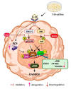
An overview of the process by which EGCG inhibits IL-1β’s induction of uPAR expression in T24 cells. By increasing uPAR expression, IL-1β stimulates the invasion of T24 cells. Through the ERK1/2/JNK signaling pathways in T24 cells, IL-1β stimulates the transcriptional activity of AP-1 and NF-κB, which in turn promotes uPAR expression. By preventing the production of uPAR and attenuating the (ERK1/2, JNK)/AP-1 and (ERK1/2, JNK)/ NF-κB signaling pathways, EGCG prevents the invasion of T24 cells that are triggered by IL-1β.

References

    1. Seebacher N.A., Stacy A.E., Porter G.M., Merlot A.M. Clinical development of targeted and immune based anti-cancer therapies. J. Exp. Clin. Cancer Res. 2019;38:156. doi: 10.1186/s13046-019-1094-2. - DOI - PMC - PubMed
    1. Khoi P.N., Li S., Thuan U.T., Sah D.K., Kang T.W., Nguyen T.T., Lian S., Xia Y., Jung Y.D. Lysophosphatidic Acid Upregulates Recepteur D’origine Nantais Expression and Cell Invasion via Egr-1, AP-1, and NF-kappaB Signaling in Bladder Carcinoma Cells. Int. J. Mol. Sci. 2020;21:304. doi: 10.3390/ijms21010304. - DOI - PMC - PubMed
    1. DeGeorge K.C., Holt H.R., Hodges S.C. Bladder Cancer: Diagnosis and Treatment. Am. Fam. Physician. 2017;96:507–514. - PubMed
    1. Ravindranathan D., Master V.A., Bilen M.A. Inflammatory Markers in Cancer Immunotherapy. Biology. 2021;10:325. doi: 10.3390/biology10040325. - DOI - PMC - PubMed
    1. Chauhan R., Trivedi V. Inflammatory markers in cancer: Potential resources. Front. Biosci. 2020;12:1–24. doi: 10.2741/s537. - DOI - PubMed

MeSH terms

Substances