. 2022 Nov 17;23(22):14232.
doi: 10.3390/ijms232214232.

The Lack of STING Impairs the MHC-I Dependent Antigen Presentation and JAK/STAT Signaling in Murine Macrophages

Affiliations

The Lack of STING Impairs the MHC-I Dependent Antigen Presentation and JAK/STAT Signaling in Murine Macrophages

Carmen Caiazza et al. Int J Mol Sci. .

Abstract

STING is a transmembrane ER resident protein that was initially described as a regulator of innate immune response triggered by viral DNA and later found to be involved in a broader range of immune processes. Here, we assessed its role in the antigen presentation by generating a STING KO macrophage cell line. In the absence of STING, we observed an impaired OVA-derived SIINFEKL peptide presentation together with a decreased level of MHC-I complex on the plasma membrane, likely due to a decreased mRNA expression of β2 m light chain as no relevant alterations of the peptide-loading complex (TAPs) were found. Moreover, JAK-STAT signaling resulted in impaired STING KO cells following OVA and LPS treatments, suggesting a dampened activation of immune response. Our data revealed a new molecular role of STING in immune mechanisms that could elucidate its role in the pathogenesis of autoimmune disorders and cancer.

Keywords: JAK/STAT; STING; antigen presentation; cell biology.


Conflict of interest statement

The authors declare no conflict of interest.

Figures

Figure 1
Figure 1
MHC-I-dependent antigen presentation of ovalbumin is impaired in STING KO macrophages. (A) J774 CTRL and STING KO (1 × 10⁶) were treated with 500 μg/mL of OVA for 24 h and were stained with SIINFEKL/H-2Kb-PE and IgG-PE as a control. Each plot represents 10,000 events of a representative experiment. (B) Histogram of J774 CTRL and STING KO treated as above. (C) Percentage of SIINFEKL/H-2Kb-positive population in untreated or OVA treated cells. Values (mean ± SE, n = 5) are shown. The asterisks indicate a statistically significant difference compared to the untreated control, according to a Student’s t-test (p < 0.01).
Figure 2
Figure 2
STING KO does not affect uptake and proteolysis of ovalbumin in murine macrophages. (A) J774 CTRL and J774 STING KO cells (2 × 10⁵) were incubated in serum-free medium with 50 μg/mL of Alexa 488-conjugated ovalbumin for 30 min at 37 °C or on ice as a negative control. Nuclei were stained with DAPI. Original magnification 20x and white scale bars represent the length of 5 μm. (B) Mean fluorescence intensity (MFI) of Alexa 488-conjugated OVA was analyzed by cytofluorimeter. Values (mean ± SE, n = 3) are shown. (C) J774 CTRL and STING KO (2 × 10⁵) were pulsed in serum-free medium with 50 μg/mL of DQ-ovalbumin for 3 min and chased for 30 min at 37 °C. Continuous incubation on ice was performed as a negative control. Nuclei were stained with DAPI. The original magnification is 20× and white scale bars represent a length of 5 μm. (D) Mean fluorescence intensity (MFI) of DQ-OVA signal was measured with a cytofluorimeter. Values (mean ± SE, n = 3) are shown.
Figure 3
Figure 3
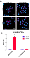
Peptide loading is impaired in STING KO macrophages. (A) J774 CTRL and STING KO cells (2 × 10⁵) were treated with 500 μg/mL of OVA for 24 h and stained with anti-MHC-I/SIINFEKL. Nuclei were stained with DAPI. Arrows indicate MHCI/SIINFEKL complexes. The original magnification is 20× and white scale bars represent a length of 5 μm. (B) A quantification analysis of MHCI/SIINFEKL dots was carried out by using a sample of 150 cells from three independent experiments. The asterisks indicate a statistically significant difference compared to the untreated control, according to a Student’s t-test (p < 0.01).
Figure 4
Figure 4
Protein expression of the components of the peptide-loading complex (TAP1, TAP2 and TPN) in control and STING KO macrophages. (A) J774 CTRL and STING KO cells (5 × 10⁶) were treated with 500 μg/mL of OVA or left untreated for the indicated time. Whole cell extracts (30 μg) were analyzed by western blot using the indicated antibodies. γ-Tubulin was included as a control for protein loading. (B) Quantifications of the protein levels of TAP1, TAP2 and TPN in CTRL (blue bars) and STING KO (red bars) were calculated by ImageJ software as fold-change relative to unstimulated, which was set to 1.0. Values (mean ± SE, n = 4) are shown. The asterisks indicate a statistically significant difference compared to the untreated control, according to a Student’s t-test (p < 0.01).
Figure 5
Figure 5
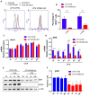
MHC-I expression on the plasma membrane and mRNA expression of H2K1 and β2 m in control and STING KO macrophages. (A) J774 CTRL and STING KO cells (1 × 10⁶) were treated with 500 μg/mL of OVA for 24 h and were stained with MHC-I-FITC and IgG-FITC as a control. Each plot represents 10,000 events of a representative experiment. (B) Percentage of MHC-I-FITC-positive population in untreated or OVA treated cells. Values (mean ± SE, n = 5) are shown. The asterisks indicate a statistically significant difference compared to the untreated control, according to a Student’s t-test (p < 0.01). (C,D) Cells (5 × 10⁶) were stimulated with 500 μg/mL of OVA for 1, 2, 4, 8, 24 h or left untreated. Total RNA was extracted and analyzed by RT-qPCR to evaluate the gene expression of H2K1 (C) and β2 m (D). Values are presented as the mean ± SD of three independent experiments. The asterisks indicate statistically significant differences between CTRL and KO cells, according to a Student’s t test (p < 0.01). (E) J774 CTRL and STING KO cells (5 × 10⁶) were treated with 500 μg/mL of OVA or left untreated for the indicated time. Whole cell extracts (30 μg) were analyzed by western blot using the indicated antibodies. γ-Tubulin was included as control for protein loading. (F) Quantifications of the protein levels of β2 m in CTRL (blue bars) and STING KO (red bars) cells were calculated by ImageJ software as fold-change relative to unstimulated, which was set to 1.0. Values (mean ± SE, n = 4) are shown.
Figure 6
Figure 6
JAK1/STAT1 activation and STAT1 expression are impaired in STING KO macrophages upon OVA or LPS treatments. (A) J774 CTRL and STING KO cells (5 × 10⁶) were treated with 500 μg/mL of OVA or left untreated for the indicated time. Aliquots of whole cell extracts (30 μg) were analyzed by western blot for the indicated proteins. γ-Tubulin was included as a control of protein loading. (B) Cells (5 × 10⁶) were stimulated with 500 μg/mL of OVA for 1, 2, 4, 8, 24 h or left untreated. Total RNA was extracted and analyzed by RT-qPCR to evaluate the gene expression of STAT1. Values (mean ± SD = 3) are indicated. The asterisks indicate statistically significant differences between CTRL and KO cells, according to a Student’s t test (p  <  0.01). (C) Murine macrophage CTRL and STING KO were stimulated with 1 μg/mL of LPS for 4 and 24 h or DMSO as a negative control. Cells were lysed and whole protein extracts were blotted and analyzed with the indicated antibodies. Blots are representative of three independent experiments.

References

    1. Huang Y.H., Liu X.Y., Du X.X., Jiang Z.F., Su X.D. The structural basis for the sensing and binding of cyclic di-gmp by sting. Nat. Struct. Mol. Biol. 2012;19:728–730. doi: 10.1038/nsmb.2333. - DOI - PubMed
    1. Srikanth S., Woo J.S., Wu B., El-Sherbiny Y.M., Leung J., Chupradit K., Rice L., Seo G.J., Calmettes G., Ramakrishna C., et al. The ca(2+) sensor stim1 regulates the type i interferon response by retaining the signaling adaptor sting at the endoplasmic reticulum. Nat. Immunol. 2019;20:152–162. doi: 10.1038/s41590-018-0287-8. - DOI - PMC - PubMed
    1. Motwani M., Pesiridis S., Fitzgerald K.A. DNA sensing by the cgas-sting pathway in health and disease. Nat. Rev. Genet. 2019;20:657–674. doi: 10.1038/s41576-019-0151-1. - DOI - PubMed
    1. Liu S., Cai X., Wu J., Cong Q., Chen X., Li T., Du F., Ren J., Wu Y.T., Grishin N.V., et al. Phosphorylation of innate immune adaptor proteins mavs, sting, and trif induces irf3 activation. Science. 2015;347:aaa2630. doi: 10.1126/science.aaa2630. - DOI - PubMed
    1. Abe T., Barber G.N. Cytosolic-DNA-mediated, sting-dependent proinflammatory gene induction necessitates canonical nf-kappab activation through tbk1. J. Virol. 2014;88:5328–5341. doi: 10.1128/JVI.00037-14. - DOI - PMC - PubMed

Substances