Review
. 2022 Nov 18;23(22):14360.
doi: 10.3390/ijms232214360.

Renal Cell Carcinoma as a Metabolic Disease: An Update on Main Pathways, Potential Biomarkers, and Therapeutic Targets

Affiliations
Review

Renal Cell Carcinoma as a Metabolic Disease: An Update on Main Pathways, Potential Biomarkers, and Therapeutic Targets

Nicola Antonio di Meo et al. Int J Mol Sci. .

Abstract

Clear cell renal cell carcinoma (ccRCC) is the most frequent histological kidney cancer subtype. Over the last decade, significant progress has been made in identifying the genetic and metabolic alterations driving ccRCC development. In particular, an integrated approach using transcriptomics, metabolomics, and lipidomics has led to a better understanding of ccRCC as a metabolic disease. The metabolic profiling of this cancer could help define and predict its behavior in terms of aggressiveness, prognosis, and therapeutic responsiveness, and would be an innovative strategy for choosing the optimal therapy for a specific patient. This review article describes the current state-of-the-art in research on ccRCC metabolic pathways and potential therapeutic applications. In addition, the clinical implication of pharmacometabolomic intervention is analyzed, which represents a new field for novel stage-related and patient-tailored strategies according to the specific susceptibility to new classes of drugs.

Keywords: Warburg effect; biomarker; metabolism; metabolomics; renal cell carcinoma.


Conflict of interest statement

The authors declare no conflict of interest.

Figures

Figure 1
Figure 1
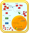
In normal cells, SIRT1 suppresses EMT and metastasis process by deacetylating SMAD4. In RCC, decreased SIRT1 activity and subsequent increased β-catenin protein levels promote an EMT-invasive phenotype. The expression levels of miR-203 in RCC cancer cells are lower than in normal cell lines. miR-203 binds toa target site and attenuates HOTAIR expression. HOTAIR enhances RCC cell migration by regulating the IGFBP2 expression. The HOTAIR–IGFBP2 axis induces glycolytic gene expression, correlating with increased proliferative and migratory capacity.
Figure 2
Figure 2
High levels of G6PD suggest that this pathway plays a major role in metabolic changes caused by RCC.
Figure 3
Figure 3
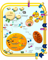
A hallmark of ccRCC is the accumulation of cholesterol, cholesterol esters, and other lipids collected in intracellular lipid droplets (LDs). LD-associated proteins, PLIN2 and HILPDA, are overexpressed in ccRCC, regulate lipid storage, and enrich lipids that contain polyunsaturated fatty-acyl side chains. Cholesterol arrives in RCC cells through direct uptake from the diet and can be acquired through receptors: very low-density lipoprotein receptor (VLDL-R) and scavenger receptor B1 (SR-B1). Once lipoprotein binds to its receptor, it forms an endosome within the membrane, which translocates into cells. Following internalization and transport into the lysosome, the cholesterol ester is hydrolyzed by lysosomal acid lipase (LAL) and upregulated in ccRCC to release the free cholesterol. To protect cells from the toxic effects of high free-cholesterol levels resulting from LAL hydrolysis, ACAT re-esterifies free cholesterol with FAs for storage inside the cell. High levels of CE in RCC tumors result from increased activity of this enzyme. Cholesterol de novo synthesis in ccRCC is reduced because of decreased levels of HMG-CoA reductase. Therefore, cholesterol accumulation in ccRCC is likely the result of increased uptake rather than excessive biosynthesis from acetate. SREBPs are transcription factors that regulate enzymes involved in cholesterol and fatty acid biosynthesis. Once inside the nucleus, they bind to the promoter regions of SREBP target genes and initiate the expression of enzymes involved in FA, TG, and cholesterol synthesis and uptake. SETD8 is stabilized by deubiquitination mediated by ubiquitin-specific protease 17 (USP17) and modulates the transcription of SREBP1 as a direct target by methylating the 20th lysine of histone-4 with enormous effects on lipogenesis. The long-chain fatty acid transferase CPT1A is decreased in ccRCC clinical samples and cell lines compared to normal samples. HIFs are responsible for inhibiting CPT1A expression, reducing FA transport into mitochondria, and rerouting FA to LDs for storage. CPT1A deficiency promotes the expression of two members of class B scavenger receptors, CD36 and SRB1, leading to increased cholesterol absorption and intracellular lipid accumulation. CD36 and SRB1 are members of class B scavenger receptors, which promote lipid and cholesterol uptake. Elongation and desaturation are the main steps of the de novo synthesis of long-chain FAs (LC-FAs). The length and degree of unsaturation are determinants of FA function and metabolic fate. Palmitate (16:0) is the main product of de novo lipogenesis. It can be elongated and desaturated through the activity of SCD1 and ELOVLs to generate additional SFAs, MUFAs, and PUFAs. ccRCC cell lines upregulate SCD1 and HIF-2α, and SCD1 had synergistic effects in sustaining cancer cell survival and migration. These effects were due to a positive feedback loop between HIF-2α and SCD1, mediated by PI3K/Akt pathway activation.
Figure 4
Figure 4
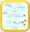
ASS1 can be negatively regulated by miR-34a-5p. The decrease in ASS1 expression favors cell proliferation. The pseudogene ASS1P3 could function as a ceRNA to regulate the expression level of its corresponding gene by competing with miR-34a-5p. A decreased level of ASS1P3 expression leads to the inhibition of ASS1 by miR-34a5p and increased cell proliferation. AR undergoes a physical interaction with ASS1P3 and regulates ASS1 expression in RCC by regulating the ceRNA activity of ASS1P3, contributing to RCC tumor progression. A decreased level of ASS1P3 expression leads to the inhibition of ASS1 by miR-34a5p and increased cell proliferation. lncRNA 00312 expression is significantly downregulated in RCC. Decreased expression of lncRNA 00312 in RCC is associated with a poorer prognosis.

References

    1. Siegel R.L., Miller K.D., Fuchs H.E., Jemal A. Cancer statistics, 2022. CA Cancer J. Clin. 2022;72:7–33. doi: 10.3322/caac.21708. - DOI - PubMed
    1. Wei J.-H., Feng Z.-H., Cao Y., Zhao H.-W., Chen Z.-H., Liao B., Wang Q., Han H., Zhang J., Xu Y.-Z., et al. Predictive value of single-nucleotide polymorphism signature for recurrence in localised renal cell carcinoma: A retrospective analysis and multicentre validation study. Lancet Oncol. 2019;20:591–600. doi: 10.1016/S1470-2045(18)30932-X. - DOI - PubMed
    1. Battaglia M., Lucarelli G. The role of renal surgery in the era of targeted therapy: The urologist’s perspective. Urologia. 2015;82:137–138. doi: 10.5301/uro.5000105. - DOI - PubMed
    1. Di Lorenzo G., De Placido S., Pagliuca M., Ferro M., Lucarelli G., Rossetti S., Bosso D., Puglia L., Pignataro P., Ascione I., et al. The evolving role of monoclonal antibodies in the treatment of patients with advanced renal cell carcinoma: A systematic review. Exp. Opin. Biol. Ther. 2016;16:1387–1401. doi: 10.1080/14712598.2016.1216964. - DOI - PubMed
    1. Tamma R., Rutigliano M., Lucarelli G., Annese T., Ruggieri S., Cascardi E., Napoli A., Battaglia M., Ribatti D. Microvascular density, macrophages, and mast cells in human clear cell renal carcinoma with and without bevacizumab treatment. Urol. Oncol. 2019;37:355.e11–355.e19. doi: 10.1016/j.urolonc.2019.01.025. - DOI - PubMed