Review
. 2022 Nov 18;23(22):14361.
doi: 10.3390/ijms232214361.

A Compilation of Study Models for Dental Pulp Regeneration

Affiliations
Review

A Compilation of Study Models for Dental Pulp Regeneration

Ella Ohlsson et al. Int J Mol Sci. .

Abstract

Efforts to heal damaged pulp tissue through tissue engineering have produced positive results in pilot trials. However, the differentiation between real regeneration and mere repair is not possible through clinical measures. Therefore, preclinical study models are still of great importance, both to gain insights into treatment outcomes on tissue and cell levels and to develop further concepts for dental pulp regeneration. This review aims at compiling information about different in vitro and in vivo ectopic, semiorthotopic, and orthotopic models. In this context, the differences between monolayer and three-dimensional cell cultures are discussed, a semiorthotopic transplantation model is introduced as an in vivo model for dental pulp regeneration, and finally, different animal models used for in vivo orthotopic investigations are presented.

Keywords: animal models; cell culture techniques; dental pulp; regeneration; regenerative endodontics; study model; tissue engineering; translational research.


Conflict of interest statement

The authors declare no conflict of interest.

Figures

Figure 1
Figure 1
Compilation of study models for dental pulp tissue engineering.
Figure 2
Figure 2
Three-dimensional cell culture models. (A) Hydrogel culture: cells are embedded in a scaffold material with a supernatant of culture medium. (B) Spheroid culture. (C) Organoid culture: cells are embedded in an extracellular matrix.
Figure 3
Figure 3
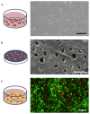
Comparison of morphologies of differently cultured cells. (A) DPSCs cultured on a tissue culture plate; scale bar of 200 µm. (B) REM image of DPSCs cultured on a dentin disk, where cells extend their processes into dentin tubules; scale bar of 10 µm. (C) Confocal laser scanning microscopy of live (green) and dead (red) DPSCs cultured in a collagen hydrogel shows the high turnover of cells; scale bar of 200 µm.
Figure 4
Figure 4
Variants of the ectopic transplantation model. (A) Dentin disk with cells seeded on top. (B) Tooth slice with cells and scaffold inserted into the pulp chamber. (C) Root fragment model with cells and scaffold inserted into the root canal.
Figure 5
Figure 5
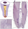
Cell-homing model. Tooth root recovered after 6 weeks of implantation into subcutaneous dorsal space of mice: (A) coronal plug, (B) dentin of root walls, (C) cells that migrated into the root canal, (D) apical reservoir of stem cells in collagen, (E) murine tissue, (F) blood vessel.
Figure 6
Figure 6
Relative tooth sizes of different species. Scale bar: 1 cm.

References

    1. Caplan D.J., Cai J., Yin G., White B.A. Root canal filled versus non-root canal filled teeth: A retrospective comparison of survival times. J. Public Health Dent. 2005;65:90–96. doi: 10.1111/j.1752-7325.2005.tb02792.x. - DOI - PubMed
    1. Yan W., Montoya C., Øilo M., Ossa A., Paranjpe A., Zhang H., Arola D.D. Contribution of root canal treatment to the fracture resistance of dentin. J. Endod. 2019;45:189–193. doi: 10.1016/j.joen.2018.10.004. - DOI - PubMed
    1. Bucchi C., Marcé-Nogué J., Galler K.M., Widbiller M. Biomechanical performance of an immature maxillary central incisor after revitalization: A finite element analysis. Int. Endod. J. 2019;52:1508–1518. doi: 10.1111/iej.13159. - DOI - PubMed
    1. Cvek M. Prognosis of luxated non-vital maxillary incisors treated with calcium hydroxide and filled with guttapercha: A retrospective clinical study. Dent. Traumatol. 1992;8:45–55. doi: 10.1111/j.1600-9657.1992.tb00228.x. - DOI - PubMed
    1. Widbiller M., Schmalz G. Endodontic regeneration: Hard shell, soft core. Odontology. 2021;109:303–312. doi: 10.1007/s10266-020-00573-1. - DOI - PMC - PubMed

LinkOut - more resources