. 2022 Nov 19;23(22):14405.
doi: 10.3390/ijms232214405.

Modular Site-Specific Conjugation of Nanobodies Using Two Co-Associating Tags

Affiliations

Modular Site-Specific Conjugation of Nanobodies Using Two Co-Associating Tags

Eric Moeglin et al. Int J Mol Sci. .

Abstract

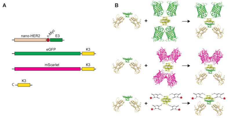
The homogeneous labeling of antibodies and their fragments is a critical step for the generation of robust probes used in immuno-detection applications. To date, numerous chemical, genetic and peptide-based site-specific coupling methods have been developed. Among these methods, co-assembling peptide-tags is one of the most straightforward and versatile solutions. Here, we describe site-specific labeling of nanobodies through the use of two co-associating peptides tags, E3 and K3, originating from the tetramerization domain of p53. These E3 and K3-tags provide a simple and robust method for associating stoichiometric amount of VHH and fluorescent probes, either fluorescent proteins or fluorochromes, at specific positions. As a proof of concept, a nanobody targeting the human epidermal growth factor receptor 2 (HER2), the nano-HER2 was genetically fused to the E3 and associated with different fluorescent K3-derivates. Entities were produced separately in Escherichia coli in soluble forms at high yields and co-assembled in vitro. These molecular probes present high binding specificity on HER2-overexpressing cells in flow-cytometry with relative binding constants in the low nanomolar range and are stable enough to stain HER2-receptor on living cells followed detection using fluorescent confocal microscopy. Altogether, our results demonstrate that the non-covalent conjugation method using these two co-associating peptides can be easily implemented for the modular engineering of molecular probes for cell immuno-staining.

Keywords: VHH; conjugation; nanobody; site-specific.


Conflict of interest statement

The authors declare no conflict of interest.

Figures

Figure 1
Figure 1
Schematic representations of the nano-HER2-E3, eGFP-K3, mScarlet-K3 and labeled-C-K3 synthetic peptides. Cartoon representing the coding sequences of each module (A) and the theoretical oligomerization states of the corresponding proteins (B) are depicted. The red star represents the Alexa-Fluor Dye.
Figure 2
Figure 2
Visualization of heterotetramer formation between the nano-HER2-E3 and either eGFP-K3, eGFP-E3 as a negative control, or synthetic K3 peptide labeled with Alexa-fluor-647 (647-C-K3) in native polyacrylamide gel electrophoresis. Constant amounts of eGFP-K3 (A) or the eGFP-E3 (B) proteins were mixed with increasing amounts of nano-HER2-E3, as indicated (* degradation product of the eGFP). On the contrary, fixed amounts of nano-HER2-E3 were mixed with increasing amounts of 647-C-K3 (C). The nano-HER2-E3 was revealed by western blot analysis with an anti-c-Myc mAb, followed by an anti-mouse mAb either IR700-labeled (A,B) or IR800-labeled (green), and both eGFP fluorescent protein and 647-C-K3 were revealed by direct fluorescence. Molar ratios between (A) nano-HER2-E3 and eGFP-K3, or (B) nano-HER2-E3 and eGFP-E3, were as follows: 1:6 (lane 3), 1:3 (lane 4), 1:2 (lane 5), 2:3 (lane 6), 5:6 (lane 7), 1:1 (lane 8) and 7:6 (lane 9). (C) Molar ratios between 647-C-K3 and nano-HER2-E3 were as follows: 0:1 (lane 1), 1:10 (lane 2), 1:5 (lane 3), 3:10 (lane 4), 3:5 (lane 5) and 1:1 (lane 6).
Figure 3
Figure 3
FRET between nano-HER2-E3-C-488 and mScarlet-K3 visualized by flow cytometry and by confocal microscopy. (A) Representation of the pair of chosen fluorophores and their respective spectra as indicated. (B) HCC1954 cells were incubated with the indicated complexes and analyzed by flow cytometry following excitation with a 488 nm laser. (C) Living fixed HCC1954 cells were incubated with the different complexes. Images were taken by confocal microscopy and analyzed with Image J. Cartoons represent the complexes. Scale bar represents 10 µm. Representative results of three independent experiments.
Figure 4
Figure 4
Determination of binding capacity of recombinant nano-HER2-E3 associated with eGFP-K3 or 488-C-K3 on HER2 overexpressing cells. (A) Cartoon representation of the heterotetramer biomolecule composed of nano-HER2-E3/eGFP-K3 binding to HER2 cell surface receptor and the positive control nano-HER2-E3-C covalently labeled with Alexa-Fluor-488 maleimide. (B) Determination of relative binding affinities of nano-HER2-E3 alone or either co-associated with eGFP-K3 or 488-C-K3 on breast cancer cells. HCC1954 cells were incubated with increasing concentrations of nano-HER2-E3-C-488, nano-HER2-E3/eGFP-K3 or nano-HER2-E3/488-C-K3, or with eGFP-K3 alone as a control. Following incubation, fluorescence was measured by flow cytometry. The relative mean fluorescence was plotted against nanobody concentration (nM), and the apparent KD value was determined using sigmoidal fitting with R software (DRC package, R Core Team GNU GPLu2 version 4.2.1 Columbia, SC, USA).

References

    1. Crauwels M., Massa S., Martin C., Betti C., Ballet S., Devoogdt N., Xavier C., Muyldermans S. Antibody Engineering. Volume 1827. Humana Press; New York, NY, USA: 2018. Site-Specific Radioactive Labeling of Nanobodies. - PubMed
    1. Massa S., Xavier C., De Vos J., Caveliers V., Lahoutte T., Muyldermans S., Devoogdt N. Site-Specific Labeling of Cysteine-Tagged Camelid Single-Domain Antibody-Fragments for Use in Molecular Imaging. Bioconjug. Chem. 2014;25:979–988. doi: 10.1021/bc500111t. - DOI - PubMed
    1. Massa S., Vikani N., Betti C., Ballet S., Vanderhaegen S., Steyaert J., Descamps B., Vanhove C., Bunschoten A., van Leeuwen F.W.B., et al. Sortase A-Mediated Site-Specific Labeling of Camelid Single-Domain Antibody-Fragments: A Versatile Strategy for Multiple Molecular Imaging Modalities. Contrast Media Mol. Imaging. 2016;11:328–339. doi: 10.1002/cmmi.1696. - DOI - PubMed
    1. Dmitriev O.Y., Lutsenko S., Muyldermans S. Nanobodies as Probes for Protein Dynamics in Vitroand in Cells. J. Biol. Chem. 2016;291:3767–3775. doi: 10.1074/jbc.R115.679811. - DOI - PMC - PubMed
    1. Pleiner T., Bates M., Trakhanov S., Lee C.T., Schliep J.E., Chug H., Böhning M., Stark H., Urlaub H., Görlich D. Nanobodies: Site-Specific Labeling for Super-Resolution Imaging, Rapid Epitope- Mapping and Native Protein Complex Isolation. Elife. 2015;4:e11349. doi: 10.7554/eLife.11349. - DOI - PMC - PubMed