

The Heart as a Target of Vasopressin and Other Cardiovascular Peptides in Health and Cardiovascular Diseases
- PMID: 36430892
- PMCID: PMC9699305
- DOI: 10.3390/ijms232214414
The Heart as a Target of Vasopressin and Other Cardiovascular Peptides in Health and Cardiovascular Diseases
Abstract
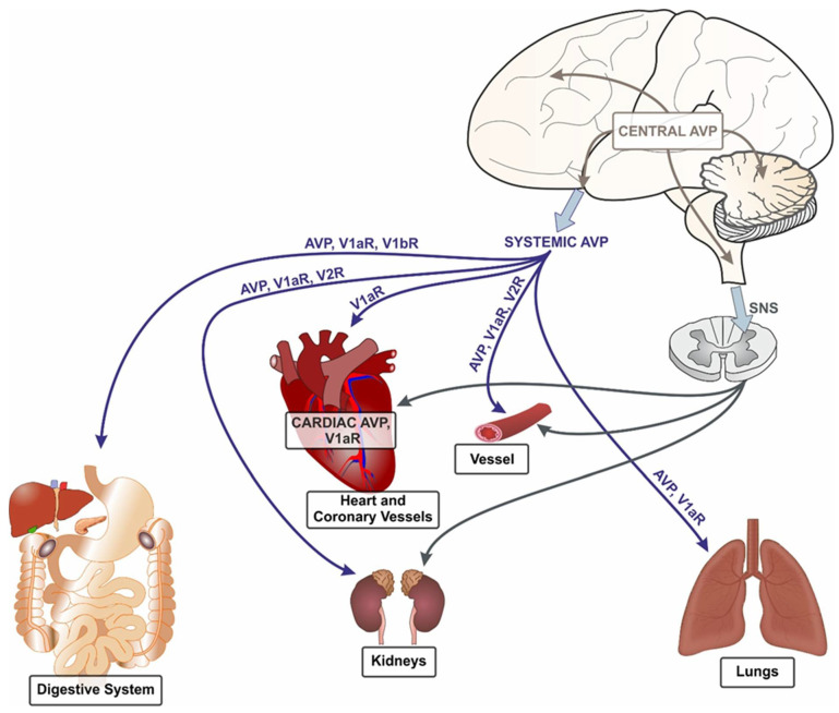
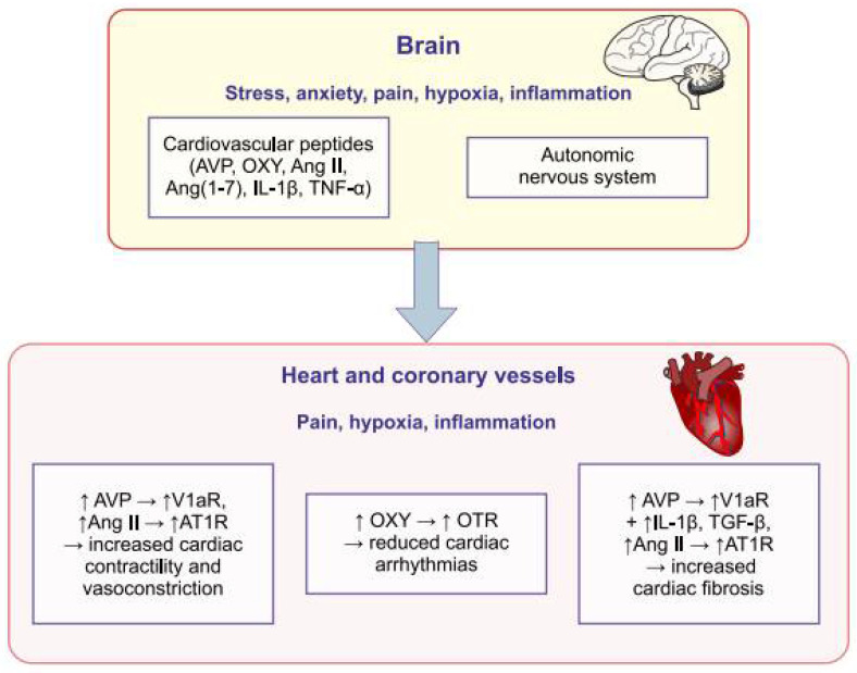
The automatism of cardiac pacemaker cells, which is tuned, is regulated by the autonomic nervous system (ANS) and multiple endocrine and paracrine factors, including cardiovascular peptides. The cardiovascular peptides (CPs) form a group of essential paracrine factors affecting the function of the heart and vessels. They may also be produced in other organs and penetrate to the heart via systemic circulation. The present review draws attention to the role of vasopressin (AVP) and some other cardiovascular peptides (angiotensins, oxytocin, cytokines) in the regulation of the cardiovascular system in health and cardiovascular diseases, especially in post-infarct heart failure, hypertension and cerebrovascular strokes. Vasopressin is synthesized mostly by the neuroendocrine cells of the hypothalamus. There is also evidence that it may be produced in the heart and lungs. The secretion of AVP and other CPs is markedly influenced by changes in blood volume and pressure, as well as by other disturbances, frequently occurring in cardiovascular diseases (hypoxia, pain, stress, inflammation). Myocardial infarction, hypertension and cardiovascular shock are associated with an increased secretion of AVP and altered responsiveness of the cardiovascular system to its action. The majority of experimental studies show that the administration of vasopressin during ventricular fibrillation and cardiac arrest improves resuscitation, however, the clinical studies do not present consisting results. Vasopressin cooperates with the autonomic nervous system (ANS), angiotensins, oxytocin and cytokines in the regulation of the cardiovascular system and its interaction with these regulators is altered during heart failure and hypertension. It is likely that the differences in interactions of AVP with ANS and other CPs have a significant impact on the responsiveness of the cardiovascular system to vasopressin in specific cardiovascular disorders.
Keywords: angiotensin; cytokines; heart failure; hypertension; hypoxia; inflammation; oxytocin; resuscitation; stress; vasopressin.
Conflict of interest statement
The author declares no conflict of interest.
Figures
Similar articles
-
Interplay of Angiotensin Peptides, Vasopressin, and Insulin in the Heart: Experimental and Clinical Evidence of Altered Interactions in Obesity and Diabetes Mellitus.Int J Mol Sci. 2024 Jan 21;25(2):1310. doi: 10.3390/ijms25021310. Int J Mol Sci. 2024. PMID: 38279313 Free PMC article. Review.
-
Complementary Role of Oxytocin and Vasopressin in Cardiovascular Regulation.Int J Mol Sci. 2021 Oct 24;22(21):11465. doi: 10.3390/ijms222111465. Int J Mol Sci. 2021. PMID: 34768894 Free PMC article. Review.
-
Cardiovascular Neuroendocrinology: Emerging Role for Neurohypophyseal Hormones in Pathophysiology.Endocrinology. 2021 Aug 1;162(8):bqab082. doi: 10.1210/endocr/bqab082. Endocrinology. 2021. PMID: 33891015 Free PMC article. Review.
-
The role of oxytocin and vasopressin in the pathophysiology of heart failure in pregnancy and in fetal and neonatal life.Am J Physiol Heart Circ Physiol. 2020 Mar 1;318(3):H639-H651. doi: 10.1152/ajpheart.00484.2019. Epub 2020 Feb 14. Am J Physiol Heart Circ Physiol. 2020. PMID: 32056469 Review.
-
Central cardiovascular effects of vasotocin, oxytocin and vasopressin in conscious rats.J Pharmacol Exp Ther. 1984 Feb;228(2):348-53. J Pharmacol Exp Ther. 1984. PMID: 6694114
Cited by
-
Unlocking the secrets of electrolytes: the prognostic value of sodium-to-chloride ratio in intensive care unit patients with myocardial infarction.BMC Cardiovasc Disord. 2024 Nov 22;24(1):664. doi: 10.1186/s12872-024-04351-7. BMC Cardiovasc Disord. 2024. PMID: 39578741 Free PMC article.
-
Aldosterone Synthase Inhibitors and Dietary Interventions: A Combined Novel Approach for Prevention and Treatment of Cardiovascular Disease.Cureus. 2023 Mar 15;15(3):e36184. doi: 10.7759/cureus.36184. eCollection 2023 Mar. Cureus. 2023. PMID: 36937127 Free PMC article. Review.
-
Molecular Interaction Between Vasopressin and Insulin in Regulation of Metabolism: Impact on Cardiovascular and Metabolic Diseases.Int J Mol Sci. 2024 Dec 11;25(24):13307. doi: 10.3390/ijms252413307. Int J Mol Sci. 2024. PMID: 39769071 Free PMC article. Review.
-
Comprehensive Insights into Mechanisms for Ventricular Remodeling in Right Heart Failure.Rev Cardiovasc Med. 2024 Nov 29;25(12):426. doi: 10.31083/j.rcm2512426. eCollection 2024 Dec. Rev Cardiovasc Med. 2024. PMID: 39742244 Free PMC article. Review.
-
Copeptin Hormone Concentrations in Dogs with Heart Disease and Relationship with Antidiuretic Hormone.Animals (Basel). 2025 Apr 1;15(7):1013. doi: 10.3390/ani15071013. Animals (Basel). 2025. PMID: 40218406 Free PMC article.
References
Publication types
MeSH terms
Substances
Grants and funding
LinkOut - more resources
Full Text Sources
Medical
Miscellaneous
