. 2022 Nov 21;23(22):14442.
doi: 10.3390/ijms232214442.

Loss of Runx1 Induces Granulosa Cell Defects and Development of Ovarian Tumors in the Mouse

Affiliations

Loss of Runx1 Induces Granulosa Cell Defects and Development of Ovarian Tumors in the Mouse

Kamiya Bridges et al. Int J Mol Sci. .

Abstract

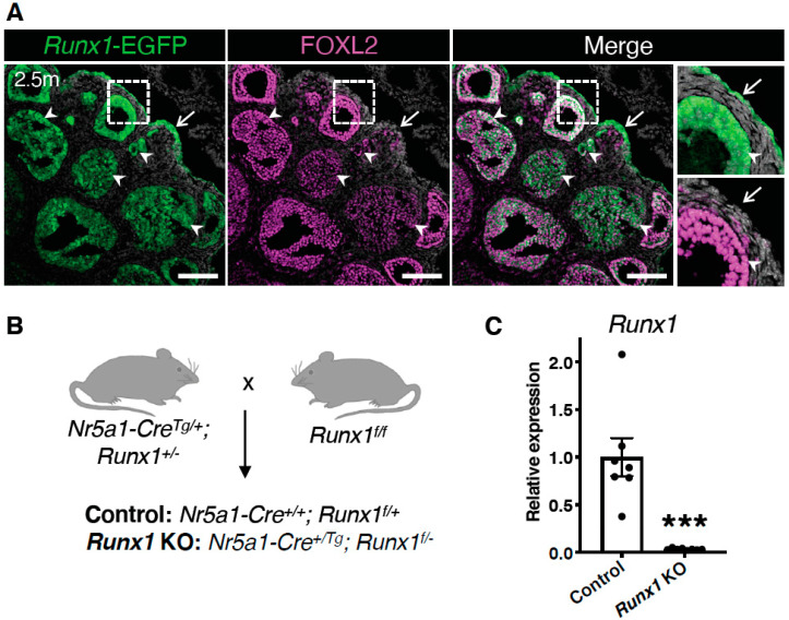
Genetic alterations of the RUNX1 gene are associated with a variety of malignancies, including female-related cancers. The role of RUNX1 as either a tumor suppressor gene or an oncogene is tissue-dependent and varies based on the cancer type. Both the amplification and deletion of the RUNX1 gene have been associated with ovarian cancer in humans. In this study, we investigated the effects of Runx1 loss on ovarian pathogenesis in mice. A conditional loss of Runx1 in the somatic cells of the ovary led to an increased prevalence of ovarian tumors in aged mice. By the age of 15 months, 27% of Runx1 knockout (KO) females developed ovarian tumors that presented characteristics of granulosa cell tumors. While ovaries from young adult mice did not display tumors, they all contained abnormal follicle-like lesions. The granulosa cells composing these follicle-like lesions were quiescent, displayed defects in differentiation and were organized in a rosette-like pattern. The RNA-sequencing analysis further revealed differentially expressed genes in Runx1 KO ovaries, including genes involved in metaplasia, ovarian cancer, epithelial cell development, tight junctions, cell-cell adhesion, and the Wnt/beta-catenin pathway. Together, this study showed that Runx1 is required for normal granulosa cell differentiation and prevention of ovarian tumor development in mice.

Keywords: RUNX1; cancer; granulosa cells; ovary; tumor.


Conflict of interest statement

The authors declare no conflict of interest.

Figures

Figure 1
Figure 1
Runx1 expression in granulosa cells and surface epithelium of adult mouse ovaries. (A) Detection of Runx1-EGFP and FOXL2 by immunofluorescence in mouse ovaries at 2.5 months of age. Grey color represents nuclei labelled with 4′,6-diamidino-2-phenylindole (DAPI). Arrows point to ovarian surface epithelium. Arrowheads point to granulosa cells. The far-right panels are higher magnifications of outlined areas. Scale bars: 250 μm. (B) Mating strategy for the generation of the control and Runx1 conditional KO mice. All Nr5a1+ somatic cells of the ovary were targeted to create a conditional knockout. (C) Relative mRNA expression of Runx1 in control and Runx1 KO ovaries at 4.5 months (control: n = 7; KO: n = 8). Values are presented as mean ± SEM; Student t-test, *** p < 0.001.
Figure 2
Figure 2
Development of ovarian tumors in aged Runx1 knockout mice. (A) Macroscopic view of control and Runx1 KO ovaries at 15 months. White dotted lines outline the ovaries. ut represents uterus. Scale bar: 2.5 mm. (B) Percentage of mice with macroscopic ovarian tumors in control (n = 27 mice) and KO mice (n = 33 mice) at 15 months. The numbers in the parenthesis are the actual numbers of animals. (C) H&E-stained sections of representative 15-month control and Runx1 KO ovaries (n = 8/genotype). Right panels are higher magnifications of outlined areas. Arrows indicate follicles; arrowheads indicate follicle-like lesions; the black asterisk indicates corpus luteum; Cy represents cyst; red asterisks indicate eosinophilic cavities. Scale bars: 300 μm. (D) Immunofluorescence for FOXL2 (magenta) and KRT8 (yellow) in control ovaries and Runx1 KO ovarian tumors (n = 4/genotype). Grey color represents nuclei labelled with DAPI. Right panels are higher magnifications of outlined areas. Arrows indicate follicles; asterisks indicate corpora lutea; cy represents cyst; T represents tumor. Scale bars: 250 μm.
Figure 3
Figure 3
Ovarian defects in young adult Runx1 knockout mice: H&E-stained sections of representative 4.5-month control (A) and Runx1 KO ovaries (B) (n = 6/genotype). Black and orange panels on the right are higher magnifications of outlined areas. Arrows indicate normal follicles; arrowheads indicate follicle-like lesions; asterisks indicate corpora lutea; cy represents cyst. Scale bars: 200 μm.
Figure 4
Figure 4
Characterization of the follicle-like lesions in Runx1 knockout mice. (A) Immunofluorescence for granulosa cell marker FOXL2 (magenta), proliferation marker KI-67 (cyan), and TUNEL cell death labeling (yellow) in control and Runx1 KO ovaries at 4.5 months. The grey color represents DAPI nuclear counterstaining. Right panels are higher magnifications of outlined areas. Arrows point to normal follicles; arrowheads point to abnormal follicles; asterisks indicate atretic follicles with TUNEL+ dying cells; yellow arrows indicate the asymmetric organization of follicle-like lesions with one layer of FOXL2 granulosa cells. Scale bars: 250 μm. (B) Immunofluorescence for granulosa cell marker AMH (magenta) with DAPI (grey) in 4.5-month control and Runx1 KO ovaries. Scale bar: 250 μm. (C) Immunofluorescence for basal membrane protein laminin (yellow) with DAPI (grey) in 4.5-month control and Runx1 KO ovaries. Right panels are single-channel outlined areas with higher magnifications. Arrowheads point to follicle-like lesions; arrows point to rosette-like structures surrounded by laminin within the lesions; scale bar: 50 μm. (D) Immunofluorescence for granulosa cell marker FOXL2 (magenta) and epithelial marker KRT8 (yellow) in 12-month control and Runx1 KO ovaries. Arrows point to normal follicles; arrowheads point to follicle-like lesions. Scale bar: 100 μm; n = 4/genotype for all immunofluorescences.
Figure 5
Figure 5
Identification of the differentially expressed genes and pathways in Runx1 knockout ovaries. (A) Heat-map of the 349 differentially expressed genes (DEGs) in Runx1 KO vs. control ovaries at 4.5 months identified by RNA-seq (n = 6/genotype). (B) Gene Ontology Biological Process analysis based on DEGs. (C) KEGG pathway analysis based on DEGs. (D) DisGeNet analysis, which identified human diseases associated with the DEGs. (E) Examples of significantly differentially expressed genes in Runx1 KO vs. control ovaries at 4.5 months. Boxplots represent the DESeq2-normalized counts from RNA-seq data. Values are presented as mean ± SEM (n = 6/genotype); ns, not significant; * p < 0.05; ** p < 0.01; *** p < 0.001.
Figure 6
Figure 6
Upregulation of genes of the Wnt signaling pathway in Runx1 knockout ovaries. (A) Examples of significantly differentially expressed genes in Runx1 KO vs. control ovaries at 4.5 months. Boxplots represent the DESeq2-normalized counts from RNA-seq data. Values are presented as mean ± SEM (n = 6/genotype); * p < 0.05; ** p < 0.01; *** p < 0.001. (B) RNA-scope for the Rspo1 probe in control and Runx1 KO ovaries at 4.5 months (n = 4/genotype). Arrowheads point to Rspo1+ follicle-like lesions. Arrows point to follicle-like lesions with little to no Rspo1 expression. Scale bars: 100 μm.

Similar articles

Cited by

References

    1. Karnezis A.N., Cho K.R., Gilks C.B., Pearce C.L., Huntsman D.G. The disparate origins of ovarian cancers: Pathogenesis and prevention strategies. Nat. Rev. Cancer. 2017;17:65–74. doi: 10.1038/nrc.2016.113. - DOI - PubMed
    1. Romero I., Bast R.C., Jr. Minireview: Human ovarian cancer: Biology, current management, and paths to personalizing therapy. Endocrinology. 2012;153:1593–1602. doi: 10.1210/en.2011-2123. - DOI - PMC - PubMed
    1. Otalora-Otalora B.A., Henriquez B., Lopez-Kleine L., Rojas A. RUNX family: Oncogenes or tumor suppressors (Review) Oncol. Rep. 2019;42:3–19. doi: 10.3892/or.2019.7149. - DOI - PMC - PubMed
    1. De Braekeleer E., Ferec C., De Braekeleer M. RUNX1 translocations in malignant hemopathies. Anticancer Res. 2009;29:1031–1037. - PubMed
    1. Dowdy C.R., Xie R., Frederick D., Hussain S., Zaidi S.K., Vradii D., Javed A., Li X., Jones S.N., Lian J.B., et al. Definitive hematopoiesis requires Runx1 C-terminal-mediated subnuclear targeting and transactivation. Hum. Mol. Genet. 2010;19:1048–1057. doi: 10.1093/hmg/ddp568. - DOI - PMC - PubMed

Substances