Review
. 2022 Nov 21;12(11):1937.
doi: 10.3390/life12111937.

Aptamer-Based Probes for Cancer Diagnostics and Treatment

Affiliations
Review

Aptamer-Based Probes for Cancer Diagnostics and Treatment

Xueqi Hu et al. Life (Basel). .

Abstract

Aptamers are single-stranded DNA or RNA oligomers that have the ability to generate unique and diverse tertiary structures that bind to cognate molecules with high specificity. In recent years, aptamer researches have witnessed a huge surge, owing to its unique properties, such as high specificity and binding affinity, low immunogenicity and toxicity, and simplicity of synthesis with negligible batch-to-batch variation. Aptamers may bind to targets, such as various cancer biomarkers, making them applicable for a wide range of cancer diagnosis and treatment. In cancer diagnostic applications, aptamers are used as molecular probes instead of antibodies. They have the potential to detect various cancer-associated biomarkers. For cancer therapeutic purposes, aptamers can serve as therapeutic or delivery agents. The chemical stabilization and modification strategies for aptamers may expand their serum half-life and shelf life. However, aptamer-based probes for cancer diagnosis and therapy still face several challenges for successful clinical translation. A deeper understanding of nucleic acid chemistry, tissue distribution, and pharmacokinetics is required in the development of aptamer-based probes. This review summarizes their application in cancer diagnostics and treatments based on different localization of target biomarkers, as well as current challenges and future prospects.

Keywords: aptamers; cancer diagnosis; cancer therapy; cell membrane biomarkers; extracellular biomarkers; intracellular biomarkers.


Conflict of interest statement

The authors declare no conflict of interest.

Figures

Figure 1
Figure 1
Schematic diagram of the application classification of aptamers in cancer diagnosis and treatment in the past decade.
Figure 2
Figure 2
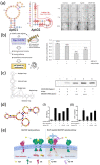
Examples of the application of aptamers as antagonists for targeted therapy of cell membrane surface biomarkers. (a) Schematic diagram of the structure of VEGFR-1 and VEGFR-2 aptamers and in vitro tube formation assay. Scale bars, 1 mm. Reprinted with permission from [132]. (b) Proposed mechanism for AS1411-induced cancer cell death and cell proliferation rate in the absence or presence of cetuximab and AS1411 aptamer. Reprinted with permission from [133]. (c) Secondary structure of SEURA-3 RNA aptamer and competitive binding assay. Reprinted with permission from [134]. (d) Secondary structure of PL1 aptamer, aptamer-mediated proliferation (I) and rescue of IFN-γ release (II) in human CD4+ T cells. Reprinted with permission from [136]. (e) Schematic diagram of bispecific aptamer targeting TfR and MET therapy. Reprinted with permission from [138].
Figure 3
Figure 3
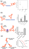
Examples of the application of aptamers as agonists for targeted therapy of cell membrane surface biomarkers. (a) Schematic of bivalent 4-1BB aptamer and its efficiency of tumor suppression. Reprinted with the permission from [141]. (b) Schematic of bivalent OX40 aptamer and OX40 activation leads to increased IFN-γ release. * p < 0.05. Reprinted with the permission from [148]. (c) Schematic of bivalent OX40 aptamer lined with streptavidin (SA)-phycoerythrin (PE) and OX40 activation leads to increased IFN-γ release. Reprinted with the permission from [142]. (d) Schematic of CD40Apt-SMG1-shRNA and its impact on overall survival. Reprinted with the permission from [143].
Figure 4
Figure 4
(a) Schematic diagram of the ATP-targeting aptamer-polymer nanogel complex and cytofluorescence image. Reprinted with permission from [229]. (b) Schematic diagram of the design of aptamer-binding alginate hydrogel targeting ATP and mouse fluorescence imaging image. Reprinted with permission from [230]. (c) Schematic diagram and cytofluorescence image of aptamer-binding AuNS nanoparticles targeting nucleolin protein on nuclear membrane. Reprinted with permission from [235]. (d) Design and cytofluorescence image of aptamer-binding mesoporous silica-coated gold nanorods. Reprinted with permission from [237].

References

    1. Cerchia L., Franciscis V.D. Targeting cancer cells with nucleic acid aptamers. Trends Biotechnol. 2010;28:517–525. doi: 10.1016/j.tibtech.2010.07.005. - DOI - PubMed
    1. Hong S., Pawel G.T., Pei R., Lu Y. Recent progress in developing fluorescent probes for imaging cell metabolites. Biomed. Mater. 2021;16:044108. doi: 10.1088/1748-605X/abfd11. - DOI - PMC - PubMed
    1. Chen H., Zhang W., Zhu G., Xie J., Chen X. Rethinking cancer nanotheranostics. Nat. Rev. Mater. 2017;2:17024. doi: 10.1038/natrevmats.2017.24. - DOI - PMC - PubMed
    1. Sun H., Zu Y. A highlight of recent advances in aptamer technology and its application. Molecules. 2015;20:11959–11980. doi: 10.3390/molecules200711959. - DOI - PMC - PubMed
    1. Mairal T., Özalp V.C., Sánchez P.L., Mir M., Katakis I., O’Sullivan C.K. Aptamers: Molecular tools for analytical applications. Anal. Bioanal. Chem. 2008;390:989–1007. doi: 10.1007/s00216-007-1346-4. - DOI - PubMed

LinkOut - more resources