Review
. 2022 Nov 8;11(22):6626.
doi: 10.3390/jcm11226626.

Embolization in Pediatric Patients: A Comprehensive Review of Indications, Procedures, and Clinical Outcomes

Affiliations
Review

Embolization in Pediatric Patients: A Comprehensive Review of Indications, Procedures, and Clinical Outcomes

Paolo Marra et al. J Clin Med. .

Abstract

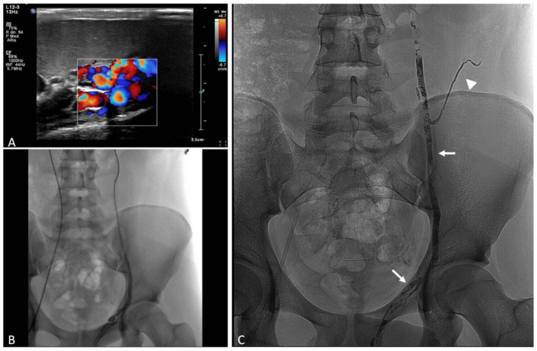
Embolization in pediatric patients encompasses a large spectrum of indications, ranging from the elective treatment of congenital diseases of the cardiovascular system to the urgent management of acute hemorrhagic conditions. In particular, the endovascular treatment of central and peripheral vascular malformations and hypervascular tumors represents a wide chapter for both congenital and acquired situations. Thanks to the progressive availability of low-profile endovascular devices and new embolic materials, the mini-invasive approach has gradually overtaken surgery. In this review, the main embolization procedures will be illustrated and discussed, with a focus on clinical indications and expected outcomes. The most recent mini-invasive techniques will be described, with hints on the cutting-edge devices and embolic materials.

Keywords: bleeding; embolization; endovascular; mini-invasive approach; pediatric.


Conflict of interest statement

The authors declare no conflict of interest.

Figures

Figure 1
Figure 1
Transcatheter coil embolization of post-traumatic splenic bleeding in a 13-year-old female. (A) Axial CTA image shows a small pseudoaneurysm (arrow) along the lateral spleen margin. (B) DSA performed through selective 4F catheterization of the celiac trunk shows several small pseudoaneurysms within the spleen parenchyma (arrows). (C) DSA image of superselective coaxial 1.9F microcatheterization of the affected spleen vessel. (D) Control DSA image after superselective multiple 4-mm coils (arrow) embolization shows disappearance of the pseudoaneurysms, with preserved opacification of the surrounding spleen parenchyma. No further bleeding occurred, nor were ischemic complications observed.
Figure 2
Figure 2
Transcatheter particle embolization of iatrogenic hepatic bleeding after percutaneous needle biopsy in a 9-year-old male. (A) Axial CTA image shows a perihepatic fluid collection (arrowheads) with contrast medium extravasation (arrow), consistent with active arterial bleeding from the site of percutaneous biopsy in the right liver lobe. (B) Selective hepatic artery digital subtraction angiography (DSA) performed with a 4F catheter shows the presence of an arterio-portal shunt with opacification of peripheral right portal branches (arrow) and contrast extravasation (arrowhead) from the lateral hepatic margin. (C) Fluoroscopic image shows superselective coaxial 1.9F microcatheterization of the artery feeding the arterio-portal shunt and the bleeding. (D) Selective hepatic artery DSA demonstrating disappearance of the bleeding after 3-mm coil (arrow) embolization.
Figure 3
Figure 3
Transcatheter coil embolization of spontaneous abdominal bleeding in a lymphoproliferative disorder of the ileum in a 10-year-old female. (A) Axial CTA image shows a large necrotic mass with hyperdense component, consistent with recent bleeding within the mesentery, with a small arterial pseudoaneurysm (arrow). (B) Selective superior mesenteric artery DSA performed with a 4F catheter shows the small pseudoaneurysm (arrow) affecting the ileocolic artery. (C) Fluoroscopic image shows contrast extravasation due to pseudoaneurysm rupture during superselective coaxial 2.7F microcatheterization of the ileocolic artery. (D) Control DSA image after superselective 3-mm coil (arrow) embolization shows disappearance of the pseudoaneurysm with no longer contrast extravasation.
Figure 4
Figure 4
Transcatheter ethylene vinyl alcohol (EVOH) copolymer embolization of a simple high-flow vascular malformation of the thigh in a 16-year-old female. (A) Magnetic Resonance Angiography T2-weighted image shows a hyperintense mass consistent with a vascular malformation (arrow). (B) Selective DSA of the deep femoral artery confirms the high-flow nature of the vascular malformation with a prevalent feeder (arrow). (C) Fluoroscopy image, acquired during EVOH embolization after superselective 2.7F coaxial microcatheterization of the lateral circumflex artery, shows capillary distribution of the embolic agent throughout the lesion. (D) Control DSA confirms the complete devascularization of the malformation; note little embolic material spread into the draining veins of the lesion (arrowheads).
Figure 5
Figure 5
Combined transarterial and transvenous transcatheter coil and plug embolization of a complex iatrogenic high-flow vascular malformation with systemic to pulmonary shunt in a 17-year-old male. A large left diaphragmatic high-flow vascular malformation developed after iatrogenic splenic infarction and abscess. (A) Reformatted coronal Computed Tomography Angiography (CTA) image shows the vascular malformation (arrows) fed by several arteries, among which intercostal, lumbar, diaphragmatic, and left gastric, draining into the left inferior pulmonary vein (arrowhead). The presence of a direct shunt with the pulmonary vein did not allow us to inject liquid embolics and particles in the afferent arteries, due to the risk of non-target cerebrovascular and systemic embolization. (B,C) Fluoroscopic images show the complete evaluation of the inflow and outflow of the malformation, realized through direct transarterial catheterization (arrow) and transeptal US-guided puncture of the heart to select the left inferior pulmonary vein (arrowheads); embolization of the arteries was performed with several metallic coils and plugs. (D) Reformatted coronal CTA image shows the plug (arrow) in the left inferior pulmonary vein (arrowhead) with reduced shunt.
Figure 6
Figure 6
Percutaneous sclero-embolization of a simple low-flow vascular malformation of the leg in a 13-year-old female. (A) B-mode Ultrasound (US) image shows a tangle of subcutaneous varicose vessels (arrow) draining into a dilated great saphenous vein (arrowhead). (B) Coronal T2-weighted Magnetic Resonance image shows an hyperintense mass (arrow) consistent with a vascular malformation. (C) DSA of the femoral axis demonstrates the low-flow nature of the lesion that has no significant arterial feeders. (D) Fluoroscopic image acquired during US-guided fine needle (arrowhead) puncture of the dilated vessels shows the distribution of the injected mixture of detergent sclerosant and ethiodized oil (to convey radiopacity); a tourniquet was tied on the thigh (not shown) to stop the saphenous flow and prevent pulmonary embolism. (E) B-mode US image shows post-procedural hyperechoic appearance with acoustic shadow of the sclerosed veins (arrow).
Figure 7
Figure 7
Transcatheter repair with coronary stent graft of a hepatic artery pseudoaneurysm after liver transplant and meso-rex surgery in a 6-year-old female. (A) Axial CTA image shows a pseudoaneurysm of the proximal tract of the transplanted hepatic artery (arrow). (B) Reformatted coronal CTA image highlights the presence of a hepatic artery stenosis (arrow), associated with the pseudoaneurysm. (C) DSA image shows selective 5F catheterization of the hepatic artery, confirming the presence of stenosis (arrow) and pseudoaneurysm (arrowhead); a 0.014” guidewire was advanced beyond the affected tract. (D) Control DSA, performed after deployment of two imbricated coronary 3.5-mm balloon expandable stent grafts, shows regular opacification of the artery and disappearance of the pseudoaneurysm.
Figure 8
Figure 8
Transcatheter repair with coronary stent graft of a hepatic artery pseudoaneurysm after liver transplant and percutaneous biliary drainage in a 9-year-old female presenting with hemobilia. (A) DSA image shows a small pseudoaneurysm (arrow) of the hepatic artery close to the percutaneous biliary drainage catheter. The common hepatic artery is thrombosed, and the proper hepatic artery is revascularized through the gastroduodenal artery, due to a previous endovascular procedure for anastomotic stenosis. (B) DSA image shows superselective coaxial 1.9F microcatheterization of the gastroduodenal artery through a 5F introducer sheath and a 5F coaxial guiding catheter placed at the origin of the superior mesenteric artery. (C) Control DSA, performed after deployment of a coronary 3-mm balloon expandable stent graft (arrow), shows regular opacification of the artery and disappearance of the pseudoaneurysm.
Figure 9
Figure 9
Percutaneous lymphangiography and sclero-embolization of chilous ascites, due to lymphatic leakage after liver transplant in a 3-year-old female. (A) Reformatted coronal Computed Tomography image shows abundant ascites. (B) Fluoroscopic image shows the ethiodized oil contrast medium injected into the groin lymph nodes spreading through the iliac lymphatics. (C) Abdominal X-rays lateral view shows extravasation (arrowheads) of ethiodized oil confirming the lymphatic leakage. (D) X-rays frontal panoramic view shows partial opacification of the lymphatic ducts up to the thoracic duct (arrow). After percutaneous lymphangiography the ascites progressively reduced up to complete resolution in few weeks.
Figure 10
Figure 10
Transcatheter plug occlusion of a congenital intrahepatic porto-systemic shunt in a 17-year-old female. (A) CDUS image shows an enlarged middle hepatic vein aberrantly communicating with the left portal vein branch. (B) Magnetic Resonance Angiography image offers a panoramic view of the aberrant porto-systemic shunt, useful to plan the percutaneous occlusion procedure. (C) Transjugular portal vein venography was performed through retrograde catheterization of the shunt. (D) Fluoroscopic image shows the deployment of a 24-mm vascular plug (arrow) within the shunt. (E) B-mode US image shows size normalization of the middle hepatic vein after effective shunt occlusion with the plug (arrow). The patient healed from post-prandial hyperammonemia.
Figure 11
Figure 11
Color Doppler Ultrasound findings of recurrent varicocele after surgery in a 16-year-old male. (A) B-mode US image shows high grade ectasia of the left pampiniform plexus extended to the inferior pole of testicle. (B) Color Doppler US examination shows persistent flow reversal during the Valsalva maneuver.
Figure 12
Figure 12
Transcatheter coil and glue embolization of recurrent varicocele after surgery in a 16-year-old male. (A) Selective left spermatic vein venography image shows an enlarged and refluent left spermatic vein with duplication and retrograde opacification of the pampiniform plexus. (B) Post-embolization venography image proves absence of opacification of the pampiniform plexus after distal occlusion with coils (arrow) and glue that spread into a duplicated vein (arrowhead).
Figure 13
Figure 13
Transcatheter glue embolization of varicocele in a 17-year-old male. (A) Color Doppler US image shows enlarged vessels of the pampiniform plexus with flow reversal during the Valsalva maneuver. (B) Fluoroscopic image shows selective transfemoral catheterization of the left spermatic vein up to the iliac region above the inguinal canal. (C) X-rays image shows the glue/ethiodized oil cast completely filling the left spermatic vein. Note the duplicated spermatic veins (arrows) and the collateral vessel (arrowhead) also filled with the embolic material.

Similar articles

Cited by

References

    1. Venturini M., Piacentino F., Coppola A., Bettoni V., Macchi E., De Marchi G., Curti M., Ossola C., Marra P., Palmisano A., et al. Visceral Artery Aneurysms Embolization and Other Interventional Options: State of the Art and New Perspectives. J. Clin. Med. 2021;10:2520. doi: 10.3390/jcm10112520. - DOI - PMC - PubMed
    1. Heran M.K.S., Burrill J. Vascular Pediatric Interventional Radiology. Can. Assoc. Radiol. J. 2012;63:S59–S73. doi: 10.1016/j.carj.2011.12.004. - DOI - PubMed
    1. Roebuck D.J., Barnacle A.M. Haemoptysis and bronchial artery embolization in children. Paediatr. Respir. Rev. 2008;9:95–104. doi: 10.1016/j.prrv.2007.12.003. - DOI - PubMed
    1. Guérin F., Abella S.F., McLin V., Ackermann O., Girard M., Cervoni J.P., Savale L., Hernandez-Gea V., Valla D., Hillaire S., et al. Congenital portosystemic shunts. Clin. Res. Hepatol. Gastroenterol. 2020;44:452–459. doi: 10.1016/j.clinre.2020.03.004. - DOI - PubMed
    1. Lord D.J.E., Chennapragada S.M. Embolization in Neonates and Infants. Tech. Vasc. Interv. Radiol. 2011;14:32–41. doi: 10.1053/j.tvir.2010.07.003. - DOI - PubMed

LinkOut - more resources