

Interpenetrating Low-Molecular Weight Hyaluronic Acid in Hyaluronic Acid-Based In Situ Hydrogel Scaffold for Periodontal and Oral Wound Applications
- PMID: 36433112
- PMCID: PMC9697763
- DOI: 10.3390/polym14224986
Interpenetrating Low-Molecular Weight Hyaluronic Acid in Hyaluronic Acid-Based In Situ Hydrogel Scaffold for Periodontal and Oral Wound Applications
Abstract
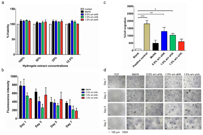
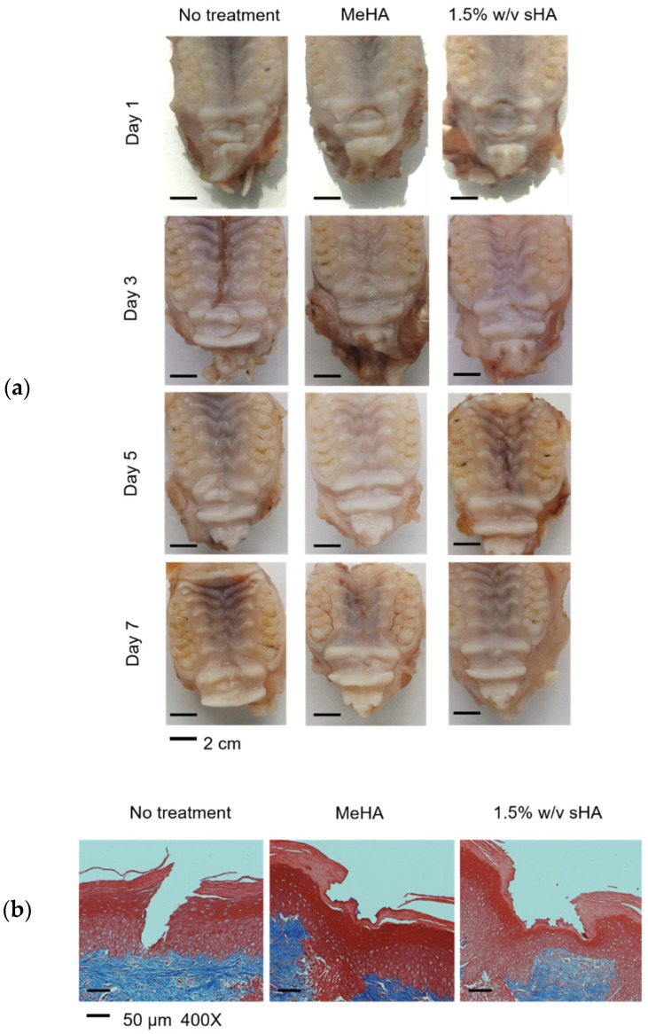
Tissues engineering has gained a lot of interest, since this approach has potential to restore lost tooth-supporting structures, which is one of the biggest challenges for periodontal treatment. In this study, we aimed to develop an in situ hydrogel that could conceivably support and promote the regeneration of lost periodontal tissues. The hydrogel was fabricated from methacrylated hyaluronic acid (MeHA). Fragment/short-chain hyaluronic acid (sHA) was incorporated in this hydrogel to encourage the bio-synergistic effects of two different molecular weights of hyaluronic acid. The physical properties of the hydrogel system, including gelation time, mechanical profile, swelling and degrading behavior, etc., were tested to assess the effect of incorporated sHA. Additionally, the biological properties of the hydrogels were performed in both in vitro and in vivo models. The results revealed that sHA slightly interfered with some behaviors of networking systems; however, the overall properties were not significantly changed compared to the base MeHA hydrogel. In addition, all hydrogel formulations were found to be compatible with oral tissues in both in vitro and in vivo models. Therefore, this HA-based hydrogel could be a promising delivery system for low molecular weight macromolecules. Further, this approach could be translated into the clinical applications for dental tissue regeneration.
Keywords: drug delivery system; hyaluronic acid; hydrogels; periodontal ligament stem cells; tissue engineering.
Conflict of interest statement
The authors declare no conflict of interest.
Figures
Similar articles
-
Structural and biological investigation of chitosan/hyaluronic acid with silanized-hydroxypropyl methylcellulose as an injectable reinforced interpenetrating network hydrogel for cartilage tissue engineering.Drug Deliv. 2021 Dec;28(1):607-619. doi: 10.1080/10717544.2021.1895906. Drug Deliv. 2021. PMID: 33739203 Free PMC article.
-
Hyaluronic acid-fibrin interpenetrating double network hydrogel prepared in situ by orthogonal disulfide cross-linking reaction for biomedical applications.Acta Biomater. 2016 Jul 1;38:23-32. doi: 10.1016/j.actbio.2016.04.041. Epub 2016 Apr 28. Acta Biomater. 2016. PMID: 27134013
-
Engineering Adhesive and Antimicrobial Hyaluronic Acid/Elastin-like Polypeptide Hybrid Hydrogels for Tissue Engineering Applications.ACS Biomater Sci Eng. 2018 Jul 9;4(7):2528-2540. doi: 10.1021/acsbiomaterials.8b00408. Epub 2018 May 16. ACS Biomater Sci Eng. 2018. PMID: 33435116 Free PMC article.
-
Interpenetrating Polymer Networks polysaccharide hydrogels for drug delivery and tissue engineering.Adv Drug Deliv Rev. 2013 Aug;65(9):1172-87. doi: 10.1016/j.addr.2013.04.002. Epub 2013 Apr 17. Adv Drug Deliv Rev. 2013. PMID: 23603210 Review.
-
Hydrogel-Based Biomaterial as a Scaffold for Gingival Regeneration: A Systematic Review of In Vitro Studies.Polymers (Basel). 2023 Jun 6;15(12):2591. doi: 10.3390/polym15122591. Polymers (Basel). 2023. PMID: 37376237 Free PMC article. Review.
Cited by
-
Hydrogels for Oral Tissue Engineering: Challenges and Opportunities.Molecules. 2023 May 7;28(9):3946. doi: 10.3390/molecules28093946. Molecules. 2023. PMID: 37175356 Free PMC article. Review.
-
Statistical optimization of hydrazone-crosslinked hyaluronic acid hydrogels for protein delivery.J Mater Chem B. 2024 Mar 6;12(10):2523-2536. doi: 10.1039/d3tb01588b. J Mater Chem B. 2024. PMID: 38344905 Free PMC article.
-
3D Bioprintable Self-Healing Hyaluronic Acid Hydrogel with Cysteamine Grafting for Tissue Engineering.Gels. 2024 Nov 28;10(12):780. doi: 10.3390/gels10120780. Gels. 2024. PMID: 39727538 Free PMC article.
-
The molecular weight of hyaluronic acid influences metabolic activity and osteogenic differentiation of periodontal ligament cells.Clin Oral Investig. 2023 Oct;27(10):5905-5911. doi: 10.1007/s00784-023-05202-z. Epub 2023 Aug 17. Clin Oral Investig. 2023. PMID: 37589747 Free PMC article.
-
An observational study of the efficacy of combined orthodontic-systemic periodontal treatment.Medicine (Baltimore). 2025 Jul 11;104(28):e43373. doi: 10.1097/MD.0000000000043373. Medicine (Baltimore). 2025. PMID: 40660512 Free PMC article. Clinical Trial.
References
-
- Sowmya S., Mony U., Jayachandran P., Reshma S., Kumar R.A., Arzate H., Nair S.V., Jayakumar R. Tri-layered nanocomposite hydrogel scaffold for the concurrent regeneration of cementum, periodontal ligament, and alveolar bone. Adv. Healthc. Mater. 2017;6:1601251. doi: 10.1002/adhm.201601251. - DOI - PubMed
Grants and funding
- CU_FRB65_hea (1)_007_32_02/Thailand Science Research and Innovation Fund Chulalongkorn University
- Ratchadapiseksompotch Fund/Chulalongkorn University
- Scholarship from the Graduate School, Chulalongkorn University to commemorate the 72nd anniversary of his Majesty King Bhumibol Adulyadej/Chulalongkorn University
- Scholarship from Second Century Fund (C2F)/Chulalongkorn University
- Grant-Supported Reseach/Asahi Glass Foundation
LinkOut - more resources
Full Text Sources
