Review
. 2022 Nov 9;22(22):8661.
doi: 10.3390/s22228661.

Graphene Incorporated Electrospun Nanofiber for Electrochemical Sensing and Biomedical Applications: A Critical Review

Affiliations
Review

Graphene Incorporated Electrospun Nanofiber for Electrochemical Sensing and Biomedical Applications: A Critical Review

Muzafar A Kanjwal et al. Sensors (Basel). .

Abstract

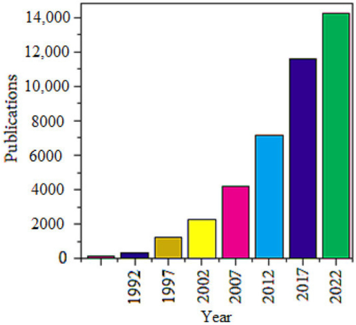
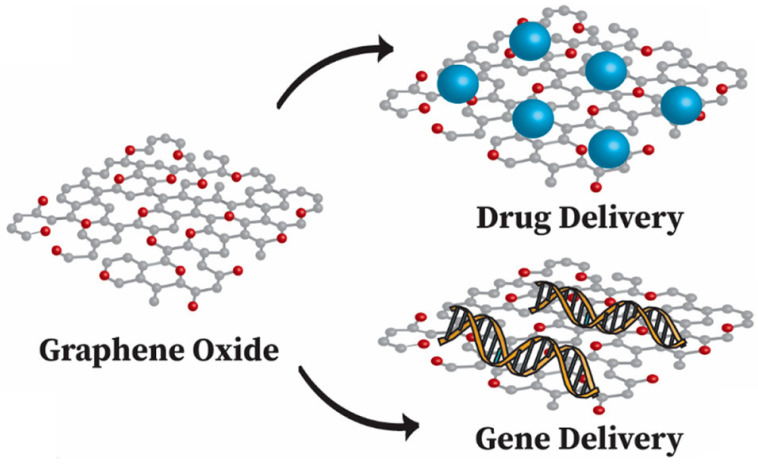
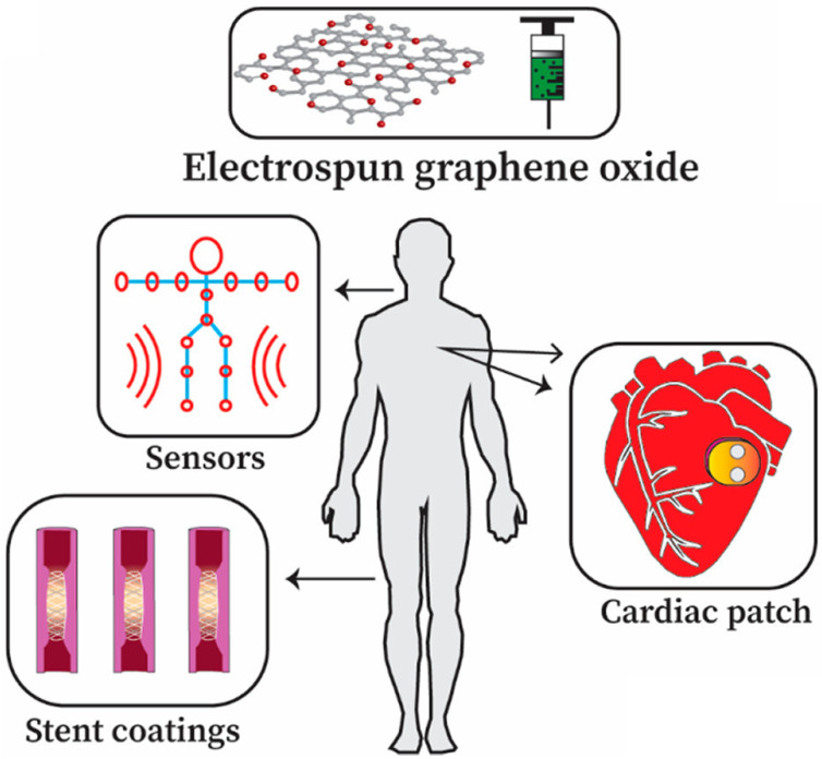
The extraordinary material graphene arrived in the fields of engineering and science to instigate a material revolution in 2004. Graphene has promptly risen as the super star due to its outstanding properties. Graphene is an allotrope of carbon and is made up of sp2-bonded carbon atoms placed in a two-dimensional honeycomb lattice. Graphite consists of stacked layers of graphene. Due to the distinctive structural features as well as excellent physico-chemical and electrical conductivity, graphene allows remarkable improvement in the performance of electrospun nanofibers (NFs), which results in the enhancement of promising applications in NF-based sensor and biomedical technologies. Electrospinning is an easy, economical, and versatile technology depending on electrostatic repulsion between the surface charges to generate fibers from the extensive list of polymeric and ceramic materials with diameters down to a few nanometers. NFs have emerged as important and attractive platform with outstanding properties for biosensing and biomedical applications, because of their excellent functional features, that include high porosity, high surface area to volume ratio, high catalytic and charge transfer, much better electrical conductivity, controllable nanofiber mat configuration, biocompatibility, and bioresorbability. The inclusion of graphene nanomaterials (GNMs) into NFs is highly desirable. Pre-processing techniques and post-processing techniques to incorporate GNMs into electrospun polymer NFs are precisely discussed. The accomplishment and the utilization of NFs containing GNMs in the electrochemical biosensing pathway for the detection of a broad range biological analytes are discussed. Graphene oxide (GO) has great importance and potential in the biomedical field and can imitate the composition of the extracellular matrix. The oxygen-rich GO is hydrophilic in nature and easily disperses in water, and assists in cell growth, drug delivery, and antimicrobial properties of electrospun nanofiber matrices. NFs containing GO for tissue engineering, drug and gene delivery, wound healing applications, and medical equipment are discussed. NFs containing GO have importance in biomedical applications, which include engineered cardiac patches, instrument coatings, and triboelectric nanogenerators (TENGs) for motion sensing applications. This review deals with graphene-based nanomaterials (GNMs) such as GO incorporated electrospun polymeric NFs for biosensing and biomedical applications, that can bridge the gap between the laboratory facility and industry.

Keywords: biomedical applications; drug delivery; electrochemical biosensors; electrospinning; electrospun nanofibers; graphene; graphene oxide; medical devices; tissue engineering; wound healing.


Conflict of interest statement

The authors declare no conflict of interest.

Figures

Figure 1
Figure 1
Publication trend of graphene from 1990–2022 Reprinted with permission from Ref. [35]. Copyright 2022, MDPI.
Figure 2
Figure 2
(A) Major fabrication methods of graphene: Top-down and bottom-up fabrication methods. Principal top-down methods include liquid-phase exfoliation and micromechanical cleavage of graphite. An additional method involves the exfoliation of initially oxidized graphite, leading to GO, which is chemically and/or thermally reduced to graphene. The bottom-up fabrication of graphene is usually performed by epitaxial growth on SiC or chemical vapor deposition, typically on Cu using small molecules, such as methane, as precursors. Reproduced with permission from [104]. Copyright Springer Nature 2020. (B) SEM (a,c,e) images and TEM images (b,d,f) of NFs (a,b), NFs-rGO-5 (c,d), and NFs-rGO-10 (e,f) with different magnifications. Reproduced with permission from [104]. Copyright 2019 Wiley.
Figure 3
Figure 3
(1): Pristine and GO-SnO2 NT preparation and gas sensor mechanism and (2) SEM images of (a) as-prepared Sn+poly (vinyl pyrrolidone) (PVP) NFs, (b,c) pristine SnO2, and (d,e) GO incorporated SnO2 NTs, (f) histogram of GO-SnO2 NT diameters. Reproduced with permission from [135]. Copyright 2019 Elsevier.
Figure 4
Figure 4
(a) Technological flow chart of the patterned STNNE. (b) FESEM image of the networked NFs. (c) FESEM image of the intersections of the NFs. (d) Optical photographs of the stretchable and transparent networked NF film. Dispersion of PU/rGO/AgNPs in NFs. (e) Raman spectra of PU/GO/AgNP nanofiber and PU/rGO/AgNP nanofiber samples with a GO:AgNP loading ratio of 1:1.25. (f) TEM images of NFs with diameters of ~290, ~484, and~933 nm. (g) Schematics of the functional groups on GO, chemical structure of polyurethane, and negative surface charges of AgNPs. GO nanosheets can be hydrogen-bonded to the PU matrix by the functional moieties of the carboxyl and hydroxyl groups. (h) Optical transmittance sheet resistance of the networked NFs for different types of NFs: rGO-coated PU, PU/rGO, PU/AgNP, and PU/rGO/AgNP NFs with that of copper nanowires, PEDOT: PSS/Zonyl/DMSO, and graphene. (i) Stress–strain curves of PU/rGO and PU/rGO/AgNP NFs. Evaluation of STNNEs under stretching conditions. (j) Resistance change (ΔR/R0) versus elongation of the PU/rGO and PU/rGO/AgNP nanofiber electrodes on PDMS substrates. (k) Resistance change (ΔR/R0) versus low strain under tensile and compressive bending of STNNEs. Reproduced with permission from [104]. Copyright 2020 Springer Nature.
Figure 5
Figure 5
Dimensionally stable anodes (DSC) (a) and thermogravimetric analysis (TGA) (b) curves of pure PS matrix and the TRG/PS nanocomposites. Reproduced with permission from [154]. Copyright 2018 Elsevier. (c) TGA curves of electrospun PVA mats mixed with GO. Reproduced with permission from [155]. Copyright 2019 American Scientific Publishers. (d) TGA curves of rGO, rGO, and AM-rGO. (e) TGA curves of electrospun PMMA/PANI/AM-rGO, PMMA/PANI/rGO, and PMMA/PANI NFs. As shown in (e), the thermal degradation temperature of PMMA/PANI/Am-rGO NFs increased to~441 °C, a magnitude higher than that of the PMMA/PANI samples at~348 °C. Both (d,e) are reproduced with permission from [156]. Copyright 2017 MDPI.
Figure 6
Figure 6
(a) Schematic presentation of electrospinning for depositing PVA/GQD onto GCE for electrochemical biosensing and catalyzing of H2O2, (b) the possible detection mechanism, (c) zeta potentials of GQDs, PVA, and PVA/GQD nanofibrous membranes at varied pH, (d) CVs of GCEs modified with PVA and PVA/GQD nanofibrous membranes, sensitivity of the biosensor at different potentials (inset), (e) CVs of the PVA/GQD nanofibrous membranes modified GCE 0.1 M PBS with different additions of H2O2. Reproduced with permission from [104]. Copyright 2020 Springer Nature.
Figure 7
Figure 7
Scheme showing the applications of graphene in different sensor streams.
Figure 8
Figure 8
Myogenin on tissue culture plate stained using immunofluorescence taken as control (a), GO sheet (b), and GOPCL electrospun fibers (c) including their quantitative analysis of the myogenin-positive nuclei (d). Immunofluorescence staining of myosin heavy chain (MHC) (eg) and the FESEM micrographs (hj) of the related samples [204]. Reproduced with permission from Ref. [210]. Copyright 2021 the American Chemical Society.
Figure 9
Figure 9
Analysis of cell adhesion and proliferation: (ac) SEM pictures of BCp-PVp/GO 5 wt % electrospun composites after (a) 7 days, (b) 14 days, and (c) 21 days. (df) Live/dead analysis [212]. Reproduced with permission from Ref. [212]. Copyright 2018 MDPI.
Figure 10
Figure 10
FE-SEM pictures of adhered and proliferated fibroblasts on PG scaffolds at magnifications of ×1.0k, ×3.0k, and ×10.0k; scale bars represent 50, 10, and 5 μm, respectively [210]. Reproduced with permission from Ref. [210]. Copyright 2021 the American Chemical Society.
Figure 11
Figure 11
Schematic of drug and gene delivery from graphene oxide Reproduced with permission from Ref. [210]. Copyright 2021 the American Chemical Society.
Figure 12
Figure 12
Release profiles of TCH in PVA/GT/TCH nanofiber (A) and PVA/GT/GO/TCH nanofiber (B) at pH 7.4. Reproduced with permission from ref [234]. Copyright 2020 Elsevier.
Figure 13
Figure 13
(a) Human lung cancer cells (A549), exposed to (b) pure DOX and (c) DOX-loaded PEO/CS/GO composite [237]. Reproduced with permission from ref [237]. Copyright 2015 Elsevier.
Figure 14
Figure 14
Schematic of how an electrospun scaffold containing GO/antimicrobial polymers may be effective in wound healing applications Reproduced with permission from Ref. [210]. Copyright 2021 the American Chemical Society.
Figure 15
Figure 15
Effect of gelatin/zinc oxide/graphene oxide composite (ZnO) on E. coli and S. aureus compared to GELP and ZGF [266]. Reproduced under the Creative Commons Attribution License with permission from ref [258]. Copyright 2017 Elsevier.
Figure 16
Figure 16
(a) Bacteriostasis of CS/PVA/GO0/Alli1 composites against various bacteria. (b) Bacteriostasis of distinctive composites against S. aureus over time. (c) The alteration of bacteriostatic circle diameter of various composites over time. Sample: a: CS/PVA/GO0/Alli0; b: CS/PVA/GO0/Alli0.5; c: CS/PVA/GO0/Alli1; d: CS/PVA/GO0/Alli2; e: CS/PVA/GO0.1/Alli2; f: CS/PVA/GO0.3/Alli2; g: CS/PVA/GO0.5/Alli2 [268]. Reproduced with permission from ref [268]. Copyright 2020 Elsevier.
Figure 17
Figure 17
Antibacterial characteristics of PCL/rGO-Ag composites. (a) S. aureus inoculum was 7.00 × 10⁵ CFU/sample. (b) E. coli O157:H7 inoculum was 6.33 × 10⁵ CFU/sample [56]. Reproduced with permission from ref [56]. Copyright 2020 Elsevier.
Figure 18
Figure 18
Potential for electrospun graphene oxide in biomaterials and medical device applications Reproduced with permission from Ref. [210]. Copyright 2021 the American Chemical Society.
Figure 19
Figure 19
RGO/TPU strain sensors used to detect various human motions. (a) Picture of stocking with RGO/TPU strain sensor attached. (b,c) Curves of response under motions of walking, running, jumping, and squatting of RGO/TPU strain sensor on the knee. (d) Picture of RGO/TPU strain sensor on the wrist of a human. (e) Curves of response of diverse bending degrees of RGO/TPU strain sensor on the wrist. (f) Curves of response on the elbow under cyclic bending of RGO/TPU strain sensor. (g) Picture of RGO/TPU strain sensor attached on the throat. (h,i) Curves of response when the wearer coughs and says “Hi”, “Hello”, and “ZZU” (of RGO/TPU strain sensor). (jl) Curves of response of RGO/TPU strain sensor on the finger, arm, and cheek. Reproduced with permission from Ref. [287]. Copyright 2016 the American Chemical Society.

References

    1. Geim A.K., Novoselov K.S. The rise of graphene. Nat. Mater. 2007;6:183–191. doi: 10.1038/nmat1849. - DOI - PubMed
    1. Chua C.K., Pumera M. Chemical reduction of graphene oxide: A synthetic chemistry viewpoint. Chem. Soc. Rev. 2014;43:291–312. doi: 10.1039/C3CS60303B. - DOI - PubMed
    1. Novoselov K.S., Colombo L., Gellert P., Schwab M., Kim K. A roadmap for graphene. Nature. 2012;490:192–200. doi: 10.1038/nature11458. - DOI - PubMed
    1. Gao L., Ni G.-X., Liu Y., Liu B., Castro Neto A.H., Loh K.P. Face-to-face transfer of wafer-scale graphene films. Nature. 2014;505:190–194. doi: 10.1038/nature12763. - DOI - PubMed
    1. Novoselov K.S., Geim A.K., Morozov S.V., Jiang D., Katsnelson M.I., Grigorieva I.V., Dubonos S.V., Firsov A.A. Two-dimensional gas of massless Dirac fermions in graphene. Nature. 2005;438:197–200. doi: 10.1038/nature04233. - DOI - PubMed

LinkOut - more resources