. 2023 Feb;22(2):e13746.
doi: 10.1111/acel.13746. Epub 2022 Nov 25.

Vascular smooth muscle cell senescence accelerates medin aggregation via small extracellular vesicle secretion and extracellular matrix reorganization

Affiliations

Vascular smooth muscle cell senescence accelerates medin aggregation via small extracellular vesicle secretion and extracellular matrix reorganization

Meredith Whitehead et al. Aging Cell. 2023 Feb.

Abstract

Vascular amyloidosis, caused when peptide monomers aggregate into insoluble amyloid, is a prevalent age-associated pathology. Aortic medial amyloid (AMA) is the most common human amyloid and is composed of medin, a 50-amino acid peptide. Emerging evidence has implicated extracellular vesicles (EVs) as mediators of pathological amyloid accumulation in the extracellular matrix (ECM). To determine the mechanisms of AMA formation with age, we explored the impact of vascular smooth muscle cell (VSMC) senescence, EV secretion, and ECM remodeling on medin accumulation. Medin was detected in EVs secreted from primary VSMCs. Small, round medin aggregates colocalized with EV markers in decellularized ECM in vitro and medin was shown on the surface of EVs deposited in the ECM. Decreasing EV secretion with an inhibitor attenuated aggregation and deposition of medin in the ECM. Medin accumulation in the aortic wall of human subjects was strongly correlated with age and VSMC senescence increased EV secretion, increased EV medin loading and triggered deposition of fibril-like medin. Proteomic analysis showed VSMC senescence induced changes in EV cargo and ECM composition, which led to enhanced EV-ECM binding and accelerated medin aggregation. Abundance of the proteoglycan, HSPG2, was increased in the senescent ECM and colocalized with EVs and medin. Isolated EVs selectively bound to HSPG2 in the ECM and its knock-down decreased formation of fibril-like medin structures. These data identify VSMC-derived EVs and HSPG2 in the ECM as key mediators of medin accumulation, contributing to age-associated AMA development.

Keywords: amyloid; extracellular matrix; extracellular vesicles; proteoglycans.


Conflict of interest statement

The authors declare that they have no conflicts of interest.

Figures
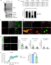
FIGURE 1
FIGURE 1
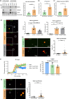
Extracellular medin deposition and aggregation is mediated by VSMC EVs. (a) Western blot of VSMC small (sEVs), medium (mEVs), large (lEVs) extracellular vesicles (EVs) and whole cell lysate (WCL). CD63 was used as a sEV marker and calnexin was used as a cell marker. (b) Slot blot of EVs and quantification showing surface localization of medin and MFG‐E8. TSG101 was used as a control which localises mainly inside the lumen of EVs, while CD63 was used as a surface protein control (n = 3 from 35‐year‐old female (35F)). (c) Immunofluorescence for medin and CD63 in decellularized ECM. Scale bar 25 μm. (d) Super‐resolution microscopy of (1) larger medin and EV aggregates, (2) individual sEVs with medin on the surface and (3) individual sEVs which do not colocalize with medin. (e) Immunofluorescent staining and quantification of ECM synthesized with or without EV secretion inhibitor 3‐O‐methylsphingomyelin (3‐OMS). Grey data points represent deposits within a field of view and black points represent average from each donor. Scale bar 25 μm, (n = 6 from 35F, 22‐year‐old male (22), 20‐year‐old male (20 M)). Unpaired Student's t test, ***p < 0.005, ****p < 0.001. (f) Thioflavin T (ThT) aggregation assay and quantification of recombinant medin peptide with or without sEVs added (n = 8–9 from 35F). Unpaired Student's t test, **p < 0.01. All data is displayed as mean ± SD and was tested for normality using Shapiro–Wilk test.
FIGURE 2
FIGURE 2
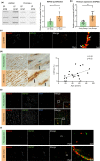
Vascular smooth muscle cell senescence enhances extracellular medin accumulation. (a) Immunohistochemistry of medin in the medial layer of aorta from human subjects. The black arrows highlight areas surrounding the nuclei which are not stained. (b) Correlation of medin staining with age (n = 25). Spearman correlation. (c) Representative Western blotting for medin and MFG‐E8 in early (EP) and late (LP) passage ECM. Fibronectin (FN) and Coomassie used as loading controls. (d) Slot blot for medin and quantification in EP ECM and LP ECM (n = 6 from 35‐year‐old female (35F), 22‐year‐old male (22 M), 20‐year‐old male (20 M)). Unpaired Student's t test, **p < 0.01. (e) Immunofluorescence of medin deposition in fibril‐like form (yellow arrow) in the LP ECM and colocalization with CD63 (white arrow) and quantification of medin deposition area and percentage of medin in fibril‐like form (n = 6 from 35F, 22 M, 20 M). Grey data points represent individual fields of view. Black data points represent average from each donor. Unpaired Student's t test, ****p < 0.001. Scale bar 25 μm. (f) Super resolution microscopy of medin in the LP ECM colocalized with CD63 (white arrows). All data is displayed as mean ± SD and was tested for normality using Shapiro–Wilk test.
FIGURE 3
FIGURE 3
Vascular smooth muscle cell senescence increases medin deposition through increased sEV secretion. (a) Western blotting and quantification for medin in extracellular vesicles (EVs) from early and late passage VSMCs (n = 4–5 from 35‐year‐old female (35F)). CD63 used as a small EV (sEV) marker. Unpaired Student's t test. (b) Secretion of sEVs from early and late passage VSMCs, quantified by flow cytometry (n = 6 from 35F, 20‐year‐old male (20 M), 22‐year‐old male (22 M)). Unpaired Student's t test, ***p < 0.005. (c) RT‐qPCR of early passage and LP VSMCs showing gene expression of neutral sphingomyelinase (SMPD3, n = 5 from 35F). Unpaired Student's t test, **p < 0.01. (d) Immunofluorescence and quantification of early passage ECM (EP ECM) and late passage ECM (LP ECM) with or without EV secretion inhibitor, 3‐O‐methylsphingomyelin (3‐OMS). Grey data points represent a field of view and black data points represent experiment averages (n = 3 from 35F). One‐way ANOVA with Tukey post hoc test, ***p < 0.005, ****p < 0.001. (e) Super‐resolution microscopy of isolated EP and LP sEVs and quantification of the area of medin on the sEVs (n = 3 from 35F). Gray data points represent individual medin‐coated sEVs and black data points represent experiment averages. Unpaired Student's t test, ****p < 0.001. (f) Thioflavin T (ThT) assay and quantification of the aggregation of recombinant medin with EP and LP sEVs added (n = 6–9 from 35F). One‐way ANOVA with Tukey's post hoc test, *p < 0.05, **p < 0.01, ****p < 0.001. (g) Immunofluorescence and quantification of medin and CD63 deposition and colocalization in the ECM. Early and late passage VSMCs were seeded on EP ECM for 5 days before being lysed. Scale bar is 25 μm. Grey data points represent individual medin deposits and black data points represent experiment averages (n = 3 from 35F). Unpaired Student's t test, ****p < 0.001. All data is displayed as mean ± SD and was tested for normality using Shapiro–Wilk test.
FIGURE 4
FIGURE 4
VSMC senescence induces changes in sEV and ECM composition. (a) Pie charts of subcellular localization of significantly changed sEV proteins which are upregulated in early passage (EP) sEVs or upregulated in late passage (LP) sEVs (n = 6 injections from 35‐year‐old female (35F) and 22‐year‐old male (22 M)). (b) Network of significantly changed proteins in LP sEVs showing the pathways which are affected. (c) Heatmap of LP vs EP sEVs with log2‐fold change (LFC) expression and enriched biological functions and pathways. (d) Heatmap of LP vs EP ECM differentially expressed proteins with LFC and enriched biological functions and pathways (n = 6 injections from 35F, 22 M, 20‐year‐old male (20 M)). (e) Protein–protein interaction network showing interactions between significantly upregulated and significantly downregulated ECM proteins in the LP ECM.
FIGURE 5
FIGURE 5
Medin and HSPG2 colocalize in the aortic medial layer and ECM. (a) Validation of the proteomics finding by slot blot and quantification of HSPG2 deposition in the early (EP) late passage (LP) ECM (n = 6 from 35‐year‐old female (35F), 20‐year‐old male (20 M) and 22‐year‐old male (22 M)). Unpaired Student's t test, *p < 0.05. (b) RT‐qPCR of HSPG2 gene expression in late passage VSMCs compared with early passage VSMCs (n = 7 from 35F, 20 M and 22 M). Unpaired Student's t test, **p < 0.01. (c) Super‐resolution microscopy of medin and HSPG2 colocalization in the LP ECM (white arrows). (d) Immunohistochemistry of extracellular HSPG2 in the aortic medial layer (black arrows). Quantification of the correlation of HSPG2 with age (n = 21). Spearman correlation. (e) Immunofluorescent staining of medin and HSPG2 deposition in human aortic medial layer. Scale bar 25 μm. (f) Super‐resolution microscopy of medin and HSPG2 colocalization in the human aorta sections. All data is displayed as mean ± SD and was tested for normality using Shapiro–Wilk test.
FIGURE 6
FIGURE 6
Increased HSPG2 deposition enhances medin aggregation. (a) Slot blot and quantification of medin and HSPG2 with HSPG2 siRNA treatment in the early passage (EP) and late passage (LP) ECM (n = 6 from 35‐year‐old female (35F)). Two‐way ANOVA with Tukey's post hoc test, *p < 0.05. (b) Immunofluorescence and quantification of medin area and fibril‐like form in LP ECM with control or siHSPG2 treatment. Gray data points represent individual fields of view and black data points represent experiment averages (n = 4 from 35F). Unpaired Student's t test, ***p < 0.005. (c) Super‐resolution microscopy of medin, CD63 and HSPG2 colocalization. (d) Immunofluorescence and quantification of sEVs from EP VSMCs (EP sEVs) or LP VSMCs (LP sEVs) binding to HSPG2 in decellularized ECM. Gray data points represent individual fields of view and black data points represent experiment averages (n = 3 from 35F). Unpaired Student's t test, *p < 0.05. (e) Super‐resolution microscopy of EP and LP sEVs bound to the ECM. Regions of HSPG2 deposition outlined in dashed white line. (f) Heatmap showing known HSPG2 binding proteins which were significantly changed in LP sEVs, compared to EP sEVs. All data are displayed as mean ± SD and were tested for normality using Shapiro–Wilk test.

Similar articles

Cited by

References

    1. Acosta, J. C. , Banito, A. , Wuestefeld, T. , Georgilis, A. , Janich, P. , Morton, J. P. , Athineos, D. , Kang, T. W. , Lasitschka, F. , Andrulis, M. , Pascual, G. , Morris, K. J. , Khan, S. , Jin, H. , Dharmalingam, G. , Snijders, A. P. , Carroll, T. , Capper, D. , Pritchard, C. , … Gil, J. (2013). A complex secretory program orchestrated by the inflammasome controls paracrine senescence. Nature Cell Biology, 15(8), 978–990. 10.1038/ncb2784 - DOI - PMC - PubMed
    1. Aguzzi, A. , & Rajendran, L. (2009). The transcellular spread of cytosolic amyloids, prions, and prionoids. Neuron, 64(6), 783–790. 10.1016/j.neuron.2009.12.016 - DOI - PubMed
    1. Alvarez‐Martinez, M. T. , Fontes, P. , Zomosa‐Signoret, V. , Arnaud, J. D. , Hingant, E. , Pujo‐Menjouet, L. , & Liautard, J. P. (2011). Dynamics of polymerization shed light on the mechanisms that lead to multiple amyloid structures of the prion protein. Biochimica et Biophysica Acta, 1814(10), 1305–1317. 10.1016/j.bbapap.2011.05.016 - DOI - PubMed
    1. Bellingham, S. A. , Guo, B. B. , Coleman, B. M. , & Hill, A. F. (2012). Exosomes: Vehicles for the transfer of toxic proteins associated with neurodegenerative diseases? Frontiers in Physiology, 3, 124. 10.3389/fphys.2012.00124 - DOI - PMC - PubMed
    1. Brown, A. M. , & Bevan, D. R. (2017). Influence of sequence and lipid type on membrane perturbation by human and rat amyloid beta‐peptide (1‐42). Archives of Biochemistry and Biophysics, 614, 1–13. 10.1016/j.abb.2016.11.006 - DOI - PubMed

Publication types

LinkOut - more resources