. 2022 Nov 24;12(1):20306.
doi: 10.1038/s41598-022-24832-z.

Visualisation of lenticulostriate arteries using contrast-enhanced time-of-flight magnetic resonance angiography at 7 Tesla

Affiliations

Visualisation of lenticulostriate arteries using contrast-enhanced time-of-flight magnetic resonance angiography at 7 Tesla

Christopher N Osuafor et al. Sci Rep. .

Abstract

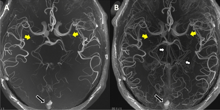
7 Tesla-field-strength (7 T) Magnetic Resonance Imaging allows the small perforating arteries in the brain to be visualised, and this modality may allow visualisation of the arterial pathology in cerebral small vessel disease. Most studies have used standard Time-of-Flight (ToF) Magnetic Resonance Angiography (MRA). Whether the use of contrast enhancement improves perforating artery visualisation at 7 T remains unclear. In a prospective study, we compared standard ToF MRA with contrast-enhanced (CE) ToF MRA at 7 T for the visualisation of the lenticulostriate arteries (LSAs). Ten patients with symptomatic lacunar stroke were recruited (mean age, SD, 64 ± 9.9 years). Visualisation was assessed using a visual rating scale administered by two independent expert readers and length of the LSAs visible. Visualisation of the LSAs was improved with CE ToF MRA. The mean Visibility and Sharpness Score was higher for CE ToF MRA over standard ToF MRA (2.55 ± 0.64 vs. 1.75 ± 0.68; P = 0.0008). The mean length of LSA visualised was significantly longer with CE ToF MRA compared to standard ToF MRA (24.4 ± 4.5 vs. 21.9 ± 4.0 mm; P = 0.01). CE ToF MRA offers improved visualisation of the LSAs over standard ToF MRA. The addition of contrast may improve the ability to visualise cerebral small vessel disease arterial pathology.


Conflict of interest statement

The authors declare no competing interests.

Figures

Figure 1
Figure 1
Qualitative analysis comparing standard ToF MRA and CE ToF MRA with boxplots and the Wilcoxon signed-rank test. For Overall Visibility and Sharpness Score, there was a statistically significant difference in favour of CE ToF MRA over standard ToF MRA. For Presence of Artefacts Score, there was no difference between standard ToF MRA and CE ToF MRA. For Extent of Venous Contamination Score, there was a difference in favour of standard ToF MRA over CE ToF MRA, however this was mild. Box plot shows X (the mean values); the 25th to 75th percentiles; the whiskers (the 5th and 95th percentiles); and the green dots (the outliers). *Statistical significance reached (P < 0.05). ToF, Time-of-Flight; MRA, Magnetic Resonance Angiography; CE, Contrast-Enhanced.
Figure 2
Figure 2
Coronal plane Maximum Intensity Projections comparing standard ToF MRA (A) and CE TOF MRA (B) for overall visibility and sharpness. The improved clarity of the LSAs in the CE ToF MRA and improved signal in areas of signal loss (yellow arrows) in the standard ToF MRA is shown. The LSAs could also be followed over a longer trajectory (yellow boxes). Also, prominence of the venous system (Choroidal and Thalamostriate Veins draining into the Internal Cerebral Veins; white arrows) is noted in the CE ToF MRA. For Overall Visibility and Sharpness Score scale, the standard ToF MRA was rated 2 (well-defined LSAs) while the CE ToF MRA was rated 3 (excellent definition of the LSAs). ToF, Time-of-Flight; MRA, Magnetic Resonance Angiography; CE, Contrast-Enhanced; LSAs, Lenticulostriate Arteries; MCA, Middle Cerebral Artery.
Figure 3
Figure 3
Axial plane Maximum Intensity Projections comparing standard ToF MRA (A) and CE ToF MRA (B) for extent of venous contamination. The prominence of the internal Cerebral Veins centrally and its tributaries (white arrows) and the Superior Sagittal Sinus (black arrows with white borders) which do not affect the visualisation of the LSAs at the MCA (yellow arrows) is shown. For Extent of Venous Contamination Score scale, this was rated 0 (no venous contamination hampering visibility) in both images. ToF, Time-of-Flight; MRA, Magnetic Resonance Angiography; CE, Contrast-Enhanced; LSAs, Lenticulostriate Arteries; MCA, Middle Cerebral Artery.

References

    1. Dichgans M, Leys D. Vascular cognitive impairment. Circ. Res. 2017;120(3):573–591. doi: 10.1161/CIRCRESAHA.116.308426. - DOI - PubMed
    1. De Cocker LJ, Lindenholz A, Zwanenburg JJ, et al. Clinical vascular imaging in the brain at 7T. Neuroimage. 2018;168:452–458. doi: 10.1016/j.neuroimage.2016.11.044. - DOI - PMC - PubMed
    1. Zwanenburg JJM, Hendrikse J, Takahara T, Visser F, Luijten PR. MR angiography of the cerebral perforating arteries with magnetization prepared anatomical reference at 7 T: Comparison with time-of-flight. J. Magn. Reson. Imaging JMRI. 2008;28(6):1519–1526. doi: 10.1002/jmri.21591. - DOI - PubMed
    1. Cho ZH, Kang CK, Han JY, et al. Observation of the lenticulostriate arteries in the human brain in vivo using 7.0T MR angiography. Stroke. 2008;39(5):1604–1606. doi: 10.1161/STROKEAHA.107.508002. - DOI - PubMed
    1. Kang CK, Park CA, Park CW, Lee YB, Cho ZH, Kim YB. Lenticulostriate arteries in chronic stroke patients visualised by 7 T magnetic resonance angiography. Int. J. Stroke. 2010;5(5):374–380. doi: 10.1111/j.1747-4949.2010.00464.x. - DOI - PubMed

Publication types