. 2023 Jun;24(2):417-434.
doi: 10.1007/s10561-022-10044-2. Epub 2022 Nov 25.

Characterization of an advanced viable bone allograft with preserved native bone-forming cells

Affiliations

Characterization of an advanced viable bone allograft with preserved native bone-forming cells

Elena Gianulis et al. Cell Tissue Bank. 2023 Jun.

Abstract

Bone grafts are widely used to successfully restore structure and function to patients with a broad range of musculoskeletal ailments and bone defects. Autogenous bone grafts are historically preferred because they theoretically contain the three essential components of bone healing (ie, osteoconductivity, osteoinductivity, and osteogenicity), but they have inherent limitations. Allograft bone derived from deceased human donors is one alternative that is also capable of providing both an osteoconductive scaffold and osteoinductive potential but, until recently, lacked the osteogenic component of bone healing. Relatively new, cellular bone allografts (CBAs) were designed to address this need by preserving viable cells. Although most commercially-available CBAs feature mesenchymal stem cells (MSCs), osteogenic differentiation is time-consuming and complex. A more advanced graft, a viable bone allograft (VBA), was thus developed to preserve lineage-committed bone-forming cells, which may be more suitable than MSCs to promote bone fusion. The purpose of this paper was to present the results of preclinical research characterizing VBA. Through a comprehensive series of in vitro and in vivo assays, the present results demonstrate that VBA in its final form is capable of providing all three essential bone remodeling properties and contains viable lineage-committed bone-forming cells, which do not elicit an immune response. The results are discussed in the context of clinical evidence published to date that further supports VBA as a potential alternative to autograft without the associated drawbacks.

Keywords: Bone graft; Bone regeneration; Bone void filler; Cellular bone allograft; Viable bone allograft; ViviGen.


Conflict of interest statement

EG, BW, DS, PG, PS, MM, and JC are employees of LifeNet Health®, the non-profit organization which processes ViviGen and paid for this study.

Figures

Fig. 1
Fig. 1
Representative photographs of VBA in its final form with either a a particulate or b fiber demineralized component
Fig. 2
Fig. 2
Representative micrographs (10 × magnification) showing a dark violet formazan-stained viable osteocytes in a three-dimensional sample of VBA following the LDH activity assay. b Following 4 to 6 weeks in growth medium, cells were observed growing from the bone chips into the medium, indicating that they remained generally active and could proliferate
Fig. 3
Fig. 3
Results of qRT-PCR analyses of osteoblast-related gene expression in cells derived from VBA (V-BC; N = 6) and human mesenchymal stem cells (hMSC; N = 3 replicates), relative to normal human osteoblast controls (NHOst; N = 3 replicates). V-BC expressed genes for osteopontin, osteocalcin, and BMP-2, a putative expression profile suggesting lineage-committed differentiation into osteoblasts
Fig. 4
Fig. 4
Representative micrographs of ICC and ICH staining for osteocalcin. For ICC (10 × magnification), a cells counterstained with hematoxylin (without osteocalcin antibody) showed presence of V-BC cells cultured from VBA, while b test samples stained positive for the putative osteoblast marker. IHC staining for osteocalcin (20 × magnification, darker red stain, denoted by arrows) in bone matrix c before and d after proprietary processing, cryopreservation, and thawing of VBA, demonstrating retention of this putative osteoblast marker in VBA’s final form
Fig. 5
Fig. 5
a Representative SEM images taken at 1 h, 1 day, or 7 days after V-BC or bmMSC were seeded onto the demineralized component of VBA (3000 × magnification). After 1 h, cells were attached to VBA (*) and demonstrated spreading and extracellular matrix deposition over the course of 7 days, demonstrating the biocompatibility and osteoconductivity of VBA. V-BC and bmMSC seeded onto the demineralized component of VBA showed significantly increased b metabolic activity, as assessed by alamarBlue assay, and c cell proliferation, as assessed by PicoGreen DNA quantification over the course of 7 days, further indicating the biocompatibility of VBA. ^p < 0.05; N = 6
Fig. 6
Fig. 6
Representative H&E staining of explants from an athymic nude mouse implanted with the demineralized component of VBA. a Merged set of images of H&E staining shows more than 50% new bone elements in the entire explant at 35 days post-implantation (4 × magnification). Expanded areas at b 4 × and c 10 × show the presence of new bone elements including new bone (*), bone marrow (&), new blood vessels (^), and chondrocytes (%) around the implanted demineralized component
Fig. 7
Fig. 7
Representative images of Alizarin Red S staining in a V-BC or b hMSC cultured in either osteogenic media (OM) or growth media (GM) for up to 7 or 14 days (10 × magnification). V-BC demonstrated Alizarin Red S staining at Day 7, which continued up to Day 14 and Day 21 (not shown), demonstrating calcium deposition and osteogenicity. No Alizarin Red S staining was detected from hMSCs at either Day 7 or Day 14. Insets show the gross observation of the entire culture well
Fig. 8
Fig. 8
Representative micrographs of IHC staining for bone marrow markers, a and b CD45 (a marker of hematopoietic cells) or c and d CD166 (a marker of MSCs) before (left panels) and after (right panels) proprietary processing, cryopreservation, and thawing of VBA, demonstrating the effective removal of bone marrow components in VBA’s final form (10 × magnification)
Fig. 9
Fig. 9
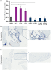
a MLR assay results demonstrating that V-BC did not induce an increase in proliferation of the HLA-mismatched PBMCs (as evidenced by no increase in BrdU incorporation compared to the PBMC only control), indicating a lack of immune cell activation. In contrast, LCs derived from the same donors as those for V-BC induced a significant increase in PBMC proliferation, with significantly greater BrdU incorporation into DNA compared to the PBMC only control, indicating immune cell activation. N = 3; *p < 0.05. Lower panels show representative images of bone matrix b before and c after VBA processing, cryopreservation, and thawing stained for MHCII surface receptors. V-BC within the bone matrix do not stain positive for MHCII, suggesting V-BC are non-immunogenic and providing an explanation of the lack of immune cell activation in the MLR assay. 10 × magnification; insets show magnified area of stained bone matrix

References

    1. Albee FH (1915) Bone graft surgery. W.B. Saunders Company, Philadelphia
    1. Alfi DM, Hassan A, East SM, Gianulis EC. Immediate mandibular reconstruction using a cellular bone allograft following tumor resection in a pediatric patient. Face. 2021 doi: 10.1177/27325016211057287. - DOI
    1. Allsopp BJ, Hunter-Smith DJ, Rozen WM. Vascularized versus nonvascularized bone grafts: What is the evidence? Clin Orthop Relat Res. 2016;474(5):1319–1327. doi: 10.1007/s11999-016-4769-4. - DOI - PMC - PubMed
    1. Alvin MD, Derakhshan A, Lubelski D, Abdullah KG, Whitmore RG, Benzel EC, et al. Cost-utility analysis of 1-and 2-level dorsal lumbar fusions with and without recombinant human bone morphogenic protein-2 at 1-year follow-up. J Spinal Disord Tech. 2016;29(1):E28–E33. doi: 10.1097/BSD.0000000000000079. - DOI - PubMed
    1. Birmingham E, Niebur G, McHugh PE (2012) Osteogenic differentiation of mesenchymal stem cells is regulated by osteocyte and osteoblast cells in a simplified bone niche. Eur Cells Mater 23:13–27. 10.22203/ecm.v023a02 - PubMed