. 2023 Mar;65(3):539-550.
doi: 10.1007/s00234-022-03088-4. Epub 2022 Nov 25.

Evaluation of the cerebrovascular reactivity in patients with Moyamoya Angiopathy by use of breath-hold fMRI: investigation of voxel-wise hemodynamic delay correction in comparison to [15O]water PET

Affiliations

Evaluation of the cerebrovascular reactivity in patients with Moyamoya Angiopathy by use of breath-hold fMRI: investigation of voxel-wise hemodynamic delay correction in comparison to [15O]water PET

Leonie Zerweck et al. Neuroradiology. 2023 Mar.

Abstract

Purpose: Patients with Moyamoya Angiopathy (MMA) require hemodynamic assessment to evaluate the risk of stroke. Hemodynamic evaluation by use of breath-hold-triggered fMRI (bh-fMRI) was proposed as a readily available alternative to the diagnostic standard [15O]water PET. Recent studies suggest voxel-wise hemodynamic delay correction in hypercapnia-triggered fMRI. The aim of this study was to evaluate the effect of delay correction of bh-fMRI in patients with MMA and to compare the results with [15O]water PET.

Methods: bh-fMRI data sets of 22 patients with MMA were evaluated without and with voxel-wise delay correction within different shift ranges and compared to the corresponding [15O]water PET data sets. The effects were evaluated combined and in subgroups of data sets with most severely impaired CVR (apparent steal phenomenon), data sets with territorial time delay, and data sets with neither steal phenomenon nor delay between vascular territories.

Results: The study revealed a high mean cross-correlation (r = 0.79, p < 0.001) between bh-fMRI and [15O]water PET. The correlation was strongly dependent on the choice of the shift range. Overall, no shift range revealed a significantly improved correlation between bh-fMRI and [15O]water PET compared to the correlation without delay correction. Delay correction within shift ranges with positive high high cutoff revealed a lower agreement between bh-fMRI and PET overall and in all subgroups.

Conclusion: Voxel-wise delay correction, in particular with shift ranges with high cutoff, should be used critically as it can lead to false-negative results in regions with impaired CVR and a lower correlation to the diagnostic standard [15O]water PET.

Keywords: Breath-hold fMRI; Cerebral perfusion reserve capacity; Cerebrovascular reactivity; Moyamoya Angiopathy; [15O]water PET.


Conflict of interest statement

The authors declare that they have no conflict of interest.

Figures

Fig. 1
Fig. 1
Volumes of interest based on the vascular territories of the anterior cerebral artery (red), the middle cerebral artery (frontal (green), temporal (yellow), and parietal (blue)), the posterior cerebral artery (pink), and the cerebellum (turquoise) separated by side [17]
Fig. 2
Fig. 2
Exemplary averaged bh-fMRI BOLD signal time-courses of each VOI of a one data set with steal phenomenon (visible in the territory of the left parietal MCA), b one data set with territorial time delay (visible in the territories of the right frontal and parietal MCA), and c one data set with neither steal phenomenon nor territorial time delay. The cerebellar signal time-courses are shown in red and the other VOIs’ signal time-courses in blue. Superimposed on each signal time-course is the mean cerebellar signal time-course in green. The duration of each averaged cycle was 69 s
Fig. 3
Fig. 3
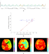
Exemplary results without and with time delay correction of one moyamoya patient. a Mean signal time-courses of each VOI without time delay correction. The cerebellar signal time-courses are shown in red and the other VOIs’ signal time-courses in blue. Superimposed on each signal time-course is the mean cerebellar signal time-course in green. b A scatter plot indicating the correlation between bh-fMRI and [15O]water PET without time delay correction (red, r = 0.81) and with exemplary time delay correction within the shift range 0–20 s (blue, r = 0.02). c Corresponding [15O]water PET map and the bh-fMRI maps without time delay correction and after time delay correction within the shift range 0–20 s. Both the scatterplots and the maps reveal a significantly weaker correlation between bh-fMRI and [15O]water PET after performing the delay correction than without delay correction
Fig. 4
Fig. 4
Patient-specific shift ranges with maximum correlation between the bh-fMRI data set and the corresponding [15O]water PET data set. The two right columns indicate the correlation between bh-fMRI and [15O]water PET without and with delay correction for each patient. The values in parentheses correspond to the correlation after the delay correction that is shown by the colored bars. Red bars indicate data sets with steal phenomenon, green bars indicate data sets with territorial delay, and blue bars represent data sets with neither steal phenomenon nor territorial delay. All correlations were significant at p < 0.05
Fig. 5
Fig. 5
Group-averaged correlation between bh-fMRI and [15O]water PET evaluating different shift ranges with variable low cutoff and high cutoff. The mean correlation between bh-fMRI and [15O]water PET is maximal at a standardized shift range with low cutoff =  − 2.25 s and high cutoff = 0 s, but not significantly higher than the correlation without delay correction (r = 0.81 vs. 0.79, p = 0.15)
Fig. 6
Fig. 6
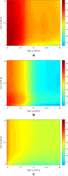
Mean correlation between bh-fMRI and [15O]water PET evaluating different shift ranges with variable low cutoff and high cutoff. The patients are sorted by subgroup affiliation: a steal phenomenon, b territorial time delay, and c neither steal phenomenon nor territorial time delay. Depending on the subgroup, the following shift ranges optima result: steal phenomenon, shift range − 1.50–0 s; territorial delay, shift range − 0.75–0 s; and neither steal phenomenon nor territorial delay, shift range − 11.25–0 s. No subgroup revealed a significant improvement in the correlation between bh-fMRI and [15O]water PET

References

    1. Suzuki J, Takaku A. Cerebrovascular “moyamoya” disease: disease showing abnormal net-like vessels in base of brain. JAMA Neurol. 1969;20:288–299. doi: 10.1001/archneur.1969.00480090076012. - DOI - PubMed
    1. Kim JS. Moyamoya disease: epidemiology, clinical features, and diagnosis. J Stroke. 2016;18:2–11. doi: 10.5853/jos.2015.01627. - DOI - PMC - PubMed
    1. Scott RM, Smith ER. Moyamoya disease and moyamoya syndrome. N Engl J Med. 2009;360:1226–1237. doi: 10.1056/NEJMra0804622. - DOI - PubMed
    1. Tarasow E, Kulakowska A, Lukasiewicz A, et al. Moyamoya disease: diagnostic imaging. Pol J Radiol. 2011;76:73–79. - PMC - PubMed
    1. Taneja K, Lu H, Welch BG, et al. Evaluation of cerebrovascular reserve in patients with cerebrovascular diseases using resting-state MRI: a feasibility study. Magn Reson Imaging. 2019;59:46–52. doi: 10.1016/j.mri.2019.03.003. - DOI - PMC - PubMed