

Primary ectopic breast carcinoma: a case report
- PMID: 36434655
- PMCID: PMC9700870
- DOI: 10.1186/s13256-022-03670-7
Primary ectopic breast carcinoma: a case report
Abstract
Background: Ectopic breast tissue is present in 2-6% of women. Ectopic mammary tissue can experience physiological changes and the same pathological processes as the eutopic breast. Ectopic breast cancer represents an uncommon condition accounting for 0.3% of all breast neoplasms, and it is most frequently located in the axilla.
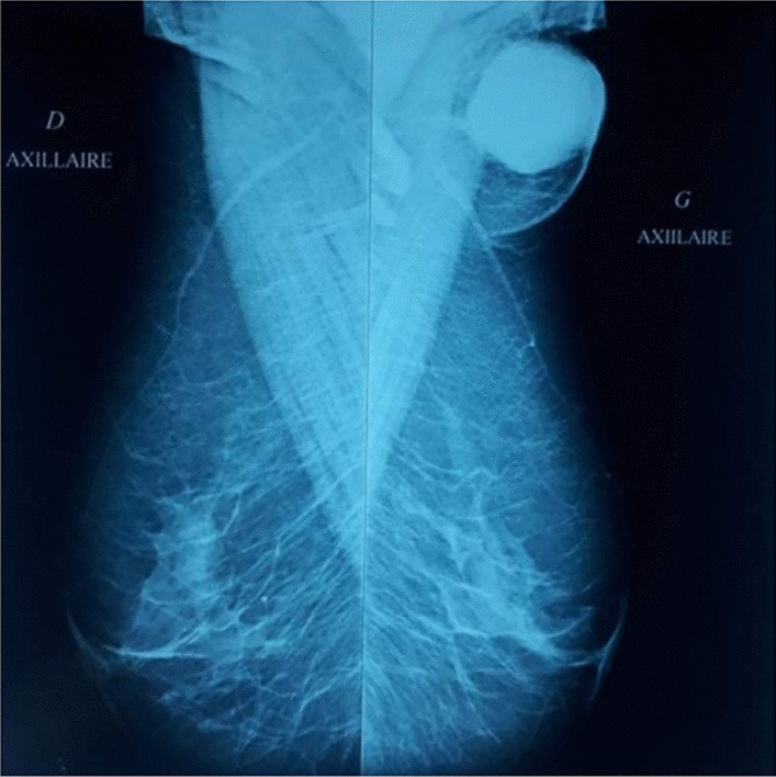
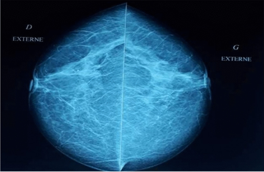
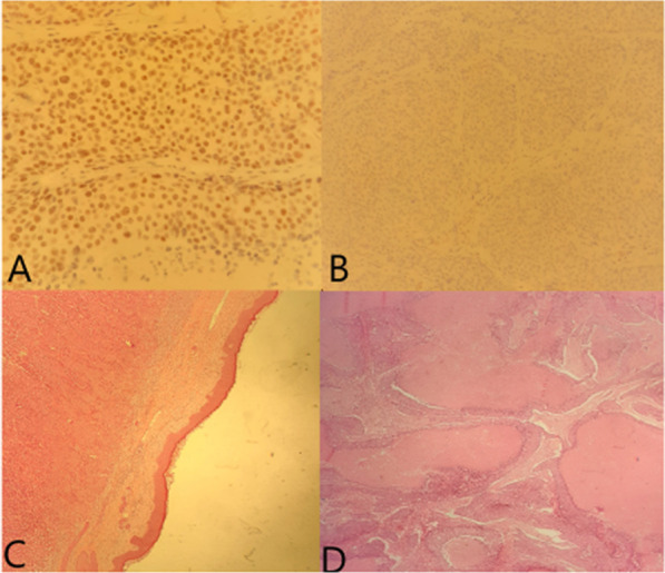
Case report: We report a rare case of a 57-year-old Tunisian woman who presented with a left-sided axillary mass evolving for about 1 month. The axillary ectopic breast tissue containing the mass was excised with axillary dissection. Pathology revealed a medullary multifocal carcinoma and metastasis was detected in two lymph nodes. She had local radiotherapy after six cycles of chemotherapy. She received herceptin therapy and hormonotherapy. After a 2-year follow-up, no evidence of local recurrence or distant metastases have been identified.
Conclusion: Ectopic breast carcinoma is a rare entity that should be the first diagnosis to be considered if an axillary lump is present in ectopic breast tissue. No particular guidelines on diagnosis and treatment are available. Therefore, physicians should be aware of this condition to avoid treatment delays. Once diagnosed, careful patient follow-up is essential because of the ambiguous natural history of this rare entity.
Keywords: Breast; Carcinoma; Case report; Ectopic; Management.
© 2022. The Author(s).
Conflict of interest statement
The authors declare that they have no competing interests.
Figures
References
Publication types
MeSH terms
LinkOut - more resources
Full Text Sources
Medical
