. 2023 Apr 6;32(8):1313-1323.
doi: 10.1093/hmg/ddac292.

Molecular basis of diseases induced by the mitochondrial DNA mutation m.9032T>C

Affiliations

Molecular basis of diseases induced by the mitochondrial DNA mutation m.9032T>C

Emilia Baranowska et al. Hum Mol Genet. .

Abstract

The mitochondrial DNA mutation m.9032T>C was previously identified in patients presenting with NARP (Neuropathy Ataxia Retinitis Pigmentosa). Their clinical features had a maternal transmission and patient's cells showed a reduced oxidative phosphorylation capacity, elevated reactive oxygen species (ROS) production and hyperpolarization of the mitochondrial inner membrane, providing evidence that m.9032T>C is truly pathogenic. This mutation leads to replacement of a highly conserved leucine residue with proline at position 169 of ATP synthase subunit a (L169P). This protein and a ring of identical c-subunits (c-ring) move protons through the mitochondrial inner membrane coupled to ATP synthesis. We herein investigated the consequences of m.9032T>C on ATP synthase in a strain of Saccharomyces cerevisiae with an equivalent mutation (L186P). The mutant enzyme assembled correctly but was mostly inactive as evidenced by a > 95% drop in the rate of mitochondrial ATP synthesis and absence of significant ATP-driven proton pumping across the mitochondrial membrane. Intragenic suppressors selected from L186P yeast restoring ATP synthase function to varying degrees (30-70%) were identified at the original mutation site (L186S) or in another position of the subunit a (H114Q, I118T). In light of atomic structures of yeast ATP synthase recently described, we conclude from these results that m.9032T>C disrupts proton conduction between the external side of the membrane and the c-ring, and that H114Q and I118T enable protons to access the c-ring through a modified pathway.


Figures

Figure 1
Figure 1
Influence of the subunit a mutations on the growth of yeast. (A) Fresh glucose cultures of the subunit a mutants and WT yeast were serially diluted and each dilution spotted on rich glucose and glycerol plates. The plates were photographed after three days of incubation at the indicated temperature. (B) Growth curves in liquid glucose and glycerol media. The shown data are representative of three independent experiments.
Figure 2
Figure 2
Influence of the L186P mutation on the stability of mitochondrial DNA. Samples from glucose cultures and L186P and WT yeasts were plated from single colonies on rich glucose plates and photographed after one-week incubation (see text for details).
Figure 3
Figure 3
Influence of the subunit a mutations on the assembly/stability and abundance of ATP synthase and Complex IV. (A) Mitochondria isolated from the subunit a mutants and WT yeast grown in rich galactose at 28°C were solubilized with digitonin (1.5 g/g protein) and separated in a 3–12% gradient polyacrylamide BN gel (200 μg of proteins/lane). The proteins were transferred to a PVDF membrane and probed with antibodies against the Atp2 (β-F1) subunit of ATP synthase. The immunological signals corresponding to dimers (V2) and monomers (V1) of ATP synthase and free F1 particles are indicated. (B) Total cellular protein extracts were separated by SDS-PAGE and then transferred to a nitrocellulose membrane and probed with antibodies against the indicated proteins. (C) The intensities of the bands in Panel B were calculated using ImageJ, normalized to porin and expressed in % of WT. Because of the large amount (72%) of ρ0 cells in cultures of the L186P mutant (where the subunit a and Cox2 cannot be synthesized), the levels of these two proteins were calculated for the part of the population (28%) that contained complete (ρ+) mtDNA (shown on the right). The standard errors were calculated from three independent experiments.
Figure 4
Figure 4
Influence of the subunit a mutations on mitochondrial membrane electrical potential. Variations in mitochondrial transmembrane potential (ΔΨ) were monitored by fluorescence quenching of Rhodamine 123 in intact mitochondria isolated from the subunit a mutants and WT yeast grown in rich galactose at 28°C. (A) ADP-driven ΔΨ consumption. (B) ATP-driven proton pumping. The additions were 25 μg/mL Rhodamine 123, 150 μg/mL mitochondrial proteins, 10 μL ethanol (EtOH), 75 μM ADP (2), 2 mM KCN, 4 μg/mL oligomycin (4), 4 μM CCCP (5) and 0.2 mM ATP (6). The shown traces are representative of at least three independent experiments.
Figure 5
Figure 5
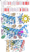
Evolutionary conservation and topology of the mutated subunit a residues. (A) Sequence alignment of helices aH3 to aH6 of subunit a from various species. The shown sequences are from Homo sapiens (H.s.), Bos taurus (B.t.), Arabidopsis thaliana (A.t.), Schizosaccharomyces pombe (S.p.), Podospora anserina (P.a.), Yarrowia lipolytica (Y.l.) and Saccharomyces cerevisiae (S.c.). Amino acid numbering in H.s. and S.c. subunits a is indicated above and below the alignments, respectively. As indicated in red, the m.9032T>C results in the replacement with proline of the leucine residue present at position 169 of H.s. subunit a. The equivalent mutation in yeast is L186P. The positions and amino acid changes induced by suppressors of L186P are indicated in yellow/brown. (B) Detail view of the p-pocket. Residues H185, E223, N100, N180 and Q230 (drawn as sticks with carbons colored in cyan) are important for moving protons from the external side of the inner membrane to the c-ring. L186 is drawn as yellow ball and stick. The H114 and I118 residues targeted by second-site suppressors are drawn as sticks with carbons colored in white. (C) Overall view from the intermembrane space (IMS) of the a/c10-ring in S.c. with the pathway (black line with arrows) along which protons are moved from the IMS to the mitochondrial matrix (19). The p- and n-pockets involved in this transfer are shown in cyan and purple background, respectively (as in panels B and D). The R176 residue of subunit a and the essential glutamate residue of subunit c (cE59) are essential for the activity of FO. (D) The π-bulge modification of aH5 induced by L186P is in yellow. Residues L173, L177, W234 and L237 (in green) belong the hydrophobic plug that separates the p- and n-site pockets. Subunits b (4), f, 8 and i/j are drawn as cartoons. For sake of clarity, the N-terminal helices of subunits a and 8 are drawn as loops. The dotted line indicates a possible pathway for protons towards the c-ring that is induced by the second-site genetic suppressors (H114Q and I118T) of L186P. Residues (E162, R169, Y241 and D244) important for moving protons along the n-pocket are in purple. (E) Side view of the p-pocket. A π-bulge due to the L186P mutation (colored in yellow), prevents the entry of protons into the p-pocket. In mutants H114Q and I118T, a bypass path between the N-terminal of subunit 8 and the C-terminal extremity of aH4 (drawn as loops) possibly allows the protons to access the bottom of the p-pocket. Mutated residues H114Q and I118T are drawn as sticks with carbons colored in yellow.

References

    1. Saraste, M. (1999) Oxidative phosphorylation at the fin de siècle. Science, 283, 1488–1493. - PubMed
    1. DiMauro, S. and Schon, E.A. (2003) Mitochondrial respiratory-chain diseases. N. Engl. J. Med., 348, 2656–2668. - PubMed
    1. Vafai, S.B. and Mootha, V.K. (2012) Mitochondrial disorders as windows into an ancient organelle. Nature, 491, 374–383. - PubMed
    1. Zeviani, M. and Carelli, V. (2007) Mitochondrial disorders. Curr. Opin. Neurol., 20, 564–571. - PubMed
    1. Wallace, D.C. (2010) Mitochondrial DNA mutations in disease and aging. Environ. Mol. Mutagen., 51, 440–450. - PubMed

Publication types

MeSH terms