. 2022 Dec 22;185(26):4999-5010.e17.
doi: 10.1016/j.cell.2022.11.009. Epub 2022 Nov 25.

Structural basis for the assembly of the type V CRISPR-associated transposon complex

Affiliations

Structural basis for the assembly of the type V CRISPR-associated transposon complex

Michael Schmitz et al. Cell. .

Abstract

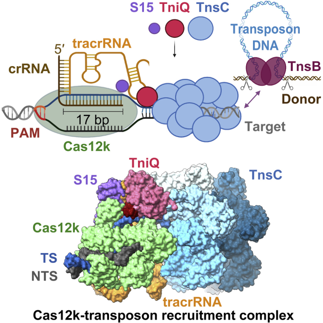
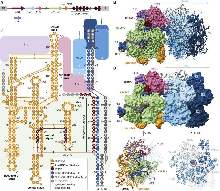
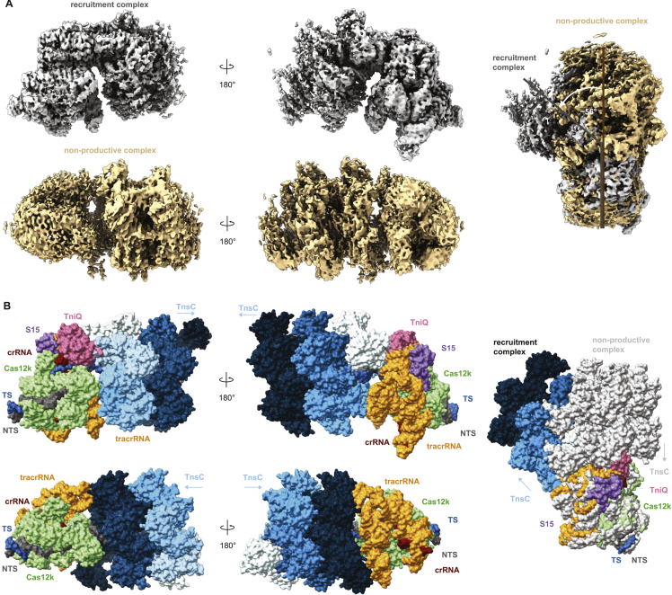
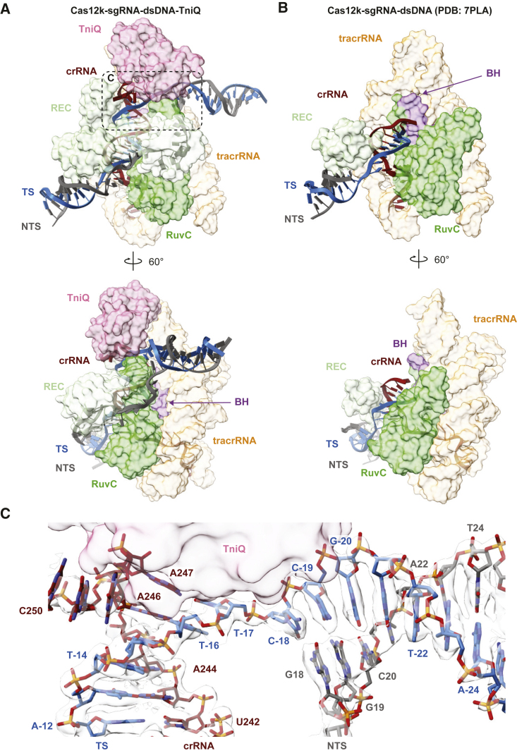
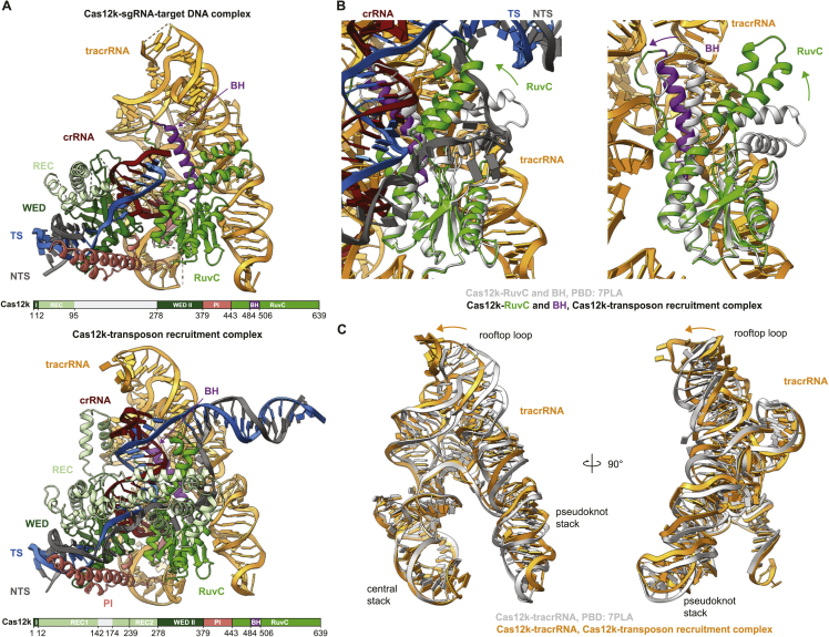
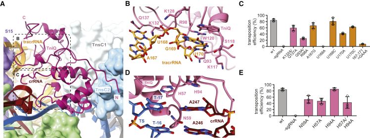
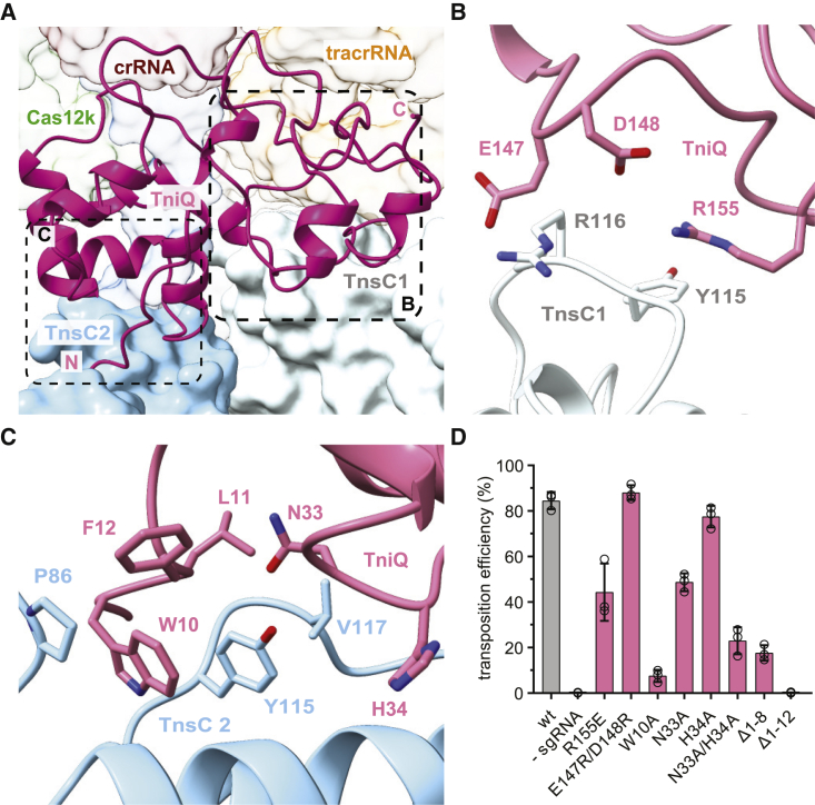
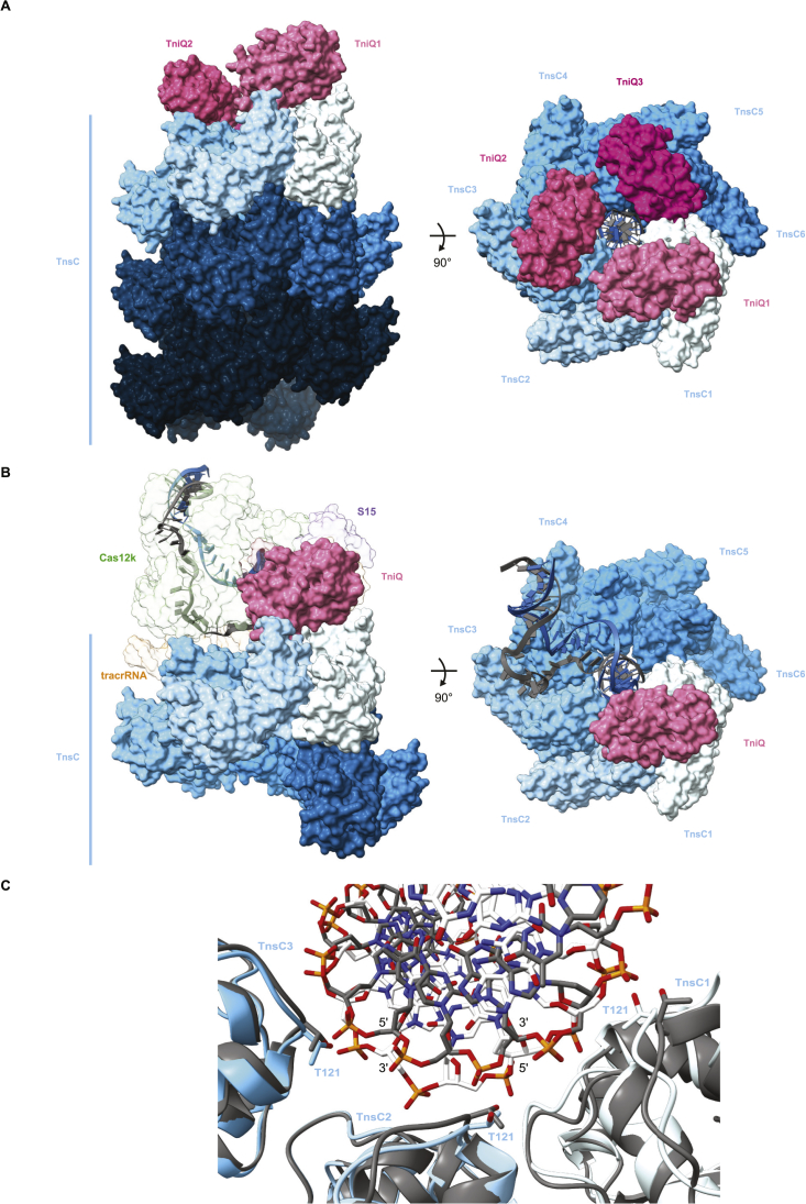
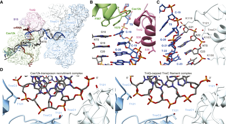
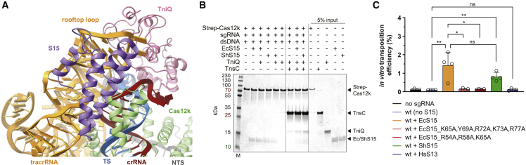
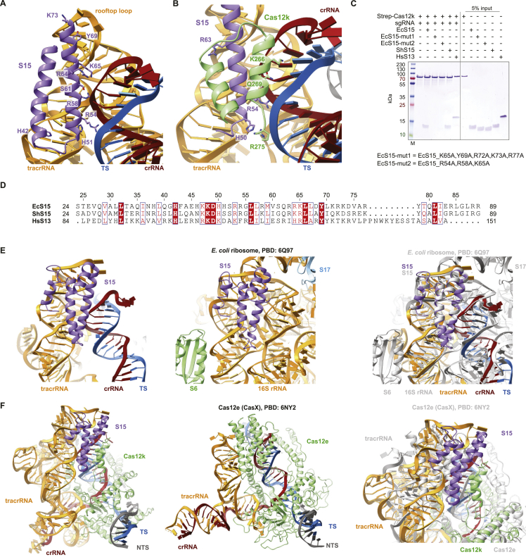
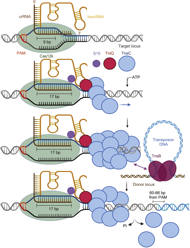
CRISPR-Cas systems have been co-opted by Tn7-like transposable elements to direct RNA-guided transposition. Type V-K CRISPR-associated transposons rely on the concerted activities of the pseudonuclease Cas12k, the AAA+ ATPase TnsC, the Zn-finger protein TniQ, and the transposase TnsB. Here we present a cryo-electron microscopic structure of a target DNA-bound Cas12k-transposon recruitment complex comprised of RNA-guided Cas12k, TniQ, a polymeric TnsC filament and, unexpectedly, the ribosomal protein S15. Complex assembly, mediated by a network of interactions involving the guide RNA, TniQ, and S15, results in R-loop completion. TniQ contacts two TnsC protomers at the Cas12k-proximal filament end, likely nucleating its polymerization. Transposition activity assays corroborate our structural findings, implying that S15 is a bona fide component of the type V crRNA-guided transposon machinery. Altogether, our work uncovers key mechanistic aspects underpinning RNA-mediated assembly of CRISPR-associated transposons to guide their development as programmable tools for site-specific insertion of large DNA payloads.

Keywords: CAST; CRISPR; CRISPR-Cas system; CRISPR-associated transposons; Cas12k; S15; TniQ; TnsB; TnsC; transposon.


Conflict of interest statement

Declaration of interests M.S., I.Q., and M.J. are named inventors on a related patent application.

Figures

None
Graphical abstract
Figure 1
Figure 1
Cryo-EM structure of the Cas12k-transposon recruitment complex (A) Schematic diagram of the type V-K ShCAST and the S15-encoding gene. LE, RE: left and right transposon ends, respectively. Spacers are represented as squares, repeats as diamonds. (B) Cryo-EM density map of the Cas12k-transposon recruitment complex. Density for nine TnsC protomers (TnsC1–TnsC9) is shown. (C) Schematic diagram of the sgRNA structure and R-loop architecture indicating interactions between nucleic acids and protein components of the complex. (D) Structural model of the Cas12k-transposon recruitment complex (surface and cartoon representations). TS, target strand; NTS, non-target strand. See also Figure S1 and Table S1.
Figure S1
Figure S1
Cryo-EM data processing workflow for the Cas12k-transposon recruitment complex, related to Figure 1 and STAR Methods (A) Cryo-EM image processing workflow for the Cas12k-transposon recruitment complex. Representative negative-stain EM and cryo-EM micrographs are shown at 98,000x and 130,000x magnifications and with 50 and 20 nm scale bars, respectively. (B) Angular distribution plotted on the density map. (C) Final cryo-EM density map colored according to local resolution. (D) Fourier Shell Correlation (FSC) of the reconstruction from two independently refined half-maps. The gold-standard cut-off (FSC = 0.143) is marked with a black dotted line. (E) Validation of the Cas12k-transposon recruitment complex atomic model.
Figure S2
Figure S2
Cryo-EM data processing workflow for the Cas12k-TnsC non-productive complex, related to Figure S3 and STAR Methods (A) Continued details of the cryo-EM image processing workflow from Figure S1. (B) Angular distribution plotted on the resulting density map. (C) Final electron density map colored according to the local resolution. (D) Fourier Shell Correlation (FSC) of the reconstruction from two independently refined half-maps. (E) Validation of the Cas12k-TnsC non-productive complex atomic model.
Figure S3
Figure S3
Structural comparison of Cas12k-transposon recruitment and Cas12k-TnsC non-productive complexes, related to Figures 1, S1, and S2 (A) Cryo-EM density maps of the Cas12k-transposon recruitment complex (top) and the Cas12k-TnsC non-productive complex (bottom). Side views and structural superpositions are shown. Proteins are shown in surface representation. The DNA in the transposon recruitment complex is bent by ∼56° relative to the non-productive complex. (B) Atomic models, shown in surface representation, of the Cas12k-transposon recruitment complex (top) and the Cas12k-TnsC non-productive complex (bottom).
Figure 2
Figure 2
R-loop completion upon complex assembly (A) Detailed views of the R-loop structure in the Cas12k-transposon recruitment complex, comprised of the crRNA portion of the single guide RNA (red cartoon backbone), the TS (blue cartoon backbone), and the NTS (dark gray cartoon backbone). Only the REC lobe, the RuvC domain, and the bridging helix (BH) of Cas12k are shown for clarity. (B) Detailed views of the R-loop structure in the Cas12k-sgRNA-target-DNA complex (PDB: 7PLA15). (C) Zoomed-in view of the PAM-distal end of the R-loop in the Cas12k-transposon recruitment complex. Density corresponding to nucleic acids is shown (contour level of 7.7 σ). TniQ is depicted as surface representation. See also Figure S4.
Figure S4
Figure S4
Structural rearrangements in Cas12k and guide RNA upon R-loop completion, related to Figure 2 (A) Structural models of the Cas12k-sgRNA-target DNA complex (PDB: 7PLA, top) and the Cas12k-transposon recruitment complex bottom), shown in the same orientation. Domain architecture of Cas12k is shown below each model. REC, recognition lobe. WED, wedge domain. PI, PAM interacting domain. BH, bridging helix. TS, target DNA strand; NTS, non-target DNA strand. (B) Structural superpositions of the RuvC and BH domains in the Cas12k-sgRNA-target DNA and Cas12k-transposon recruitment complexes. (C) Structural superposition of the tracrRNA part of the sgRNA in the Cas12k-sgRNA-target DNA (gray) and the Cas12k-transposon recruitment complex (orange).
Figure 3
Figure 3
TniQ recognizes tracrRNA and completed R-loop (A) Overview of TniQ in the Cas12k-transposon recruitment complex, depicting interfaces with the tracrRNA (orange) and the RNA:DNA heteroduplex formed by the crRNA (red) and the TS (blue). NTS is colored in dark gray. The N and C termini of TniQ are indicated. (B) Close-up view of key tracrRNA-interacting residues of TniQ. (C) Site-specific transposition activity in E. coli of ShCAST systems containing structure-based mutations in the tracrRNA or the tracrRNA-interacting interface in TniQ, as determined by droplet digital PCR (ddPCR) analysis. Data are presented as mean ± SD (n = 3 biologically independent replicates). (D) Detailed view of R-loop recognition by TniQ. (E) Site-specific transposition activity in E. coli of ShCAST systems containing structure-based mutations in the R-loop recognition interface of TniQ. Data are presented as mean ± SD (n = 3 biologically independent replicates).
Figure 4
Figure 4
TniQ contacts TnsC filament end (A) Overview of the interactions between TniQ (cartoon representation) and two TnsC protomers (TnsC1 and TnsC2, surface representation) at the Cas12k-proximal filament end. The N and C termini of TniQ are indicated. (B) Close-up view of key interactions between TniQ and TnsC1. (C) Close-up view of key interactions between TniQ and TnsC2. (D) Site-specific transposition activity in E. coli of ShCAST systems containing mutations in the TnsC-binding interface of TniQ, as determined by ddPCR analysis. Data are presented as mean ± SD (n = 3 biologically independent replicates).
Figure S5
Figure S5
Cryo-EM analysis of TniQ-capped TnsC filament, related to Figures 5 and S6 and STAR Methods (A) Cryo-EM image processing workflow for the TniQ-capped TnsC filament complex. (B) Fourier Shell Correlation (FSC) of TnsC-DNA-TniQ reconstruction from two independently refined half-maps. The gold-standard cut-off (FSC = 0.143) is marked with a blue line. (C) Final electron density map colored according to the local resolution. (D) Fourier Shell Correlation (FSC) of the reconstruction from two independently refined half-maps.
Figure S6
Figure S6
Structural comparisons of TniQ-capped TnsC filament and Cas12k-transposon recruitment complex, related to Figures 5 and S5 and Table S1 (A) Side and top views of the TniQ-capped TnsC filament. Proteins are shown in surface representation. (B) Side and top views of the Cas12k-transposon recruitment complex, with TniQ shown in the same orientation as TniQ1 in (A). In the top view, Cas12k, S15 and tracrRNA are omitted to visualize the contacts between TniQ and TnsC. (C) Structural overlay of three consecutive TnsC protomers (TnsC1-TnsC3) in the Cas12k-transposon recruitment complex (colored protomers, white DNA) and in the TniQ-capped TnsC filament (gray protomers and DNA) and the associated DNA (shown in stick representation).
Figure 5
Figure 5
TnsC assembly on PAM-distal end of R-loop DNA (A) Overview of guide-target R-loop structure within the Cas12k-transposon recruitment complex. TS (blue) and NTS (dark gray) are shown in cartoon format. Only the crRNA portion of the single-guide RNA (red) is shown. Proteins are shown in surface representation. Residues 132–254 of Cas12k and the TnsC2 and TnsC3 protomers are omitted from view for clarity. (B) Zoomed-in view of target DNA-binding residues of TniQ. (C) Zoomed-in view of the DNA-binding residues in the TnsC1 protomer. (D) Comparison of DNA binding modes of consecutive TnsC protomers (TnsC1–TnsC3) in the Cas12k-transposon recruitment complex (left) and in the TniQ-capped TnsC filament (right). See also Figure S5.
Figure 6
Figure 6
S15 promotes Cas12k-transposon recruitment complex assembly and transposition activity (A) Zoomed-in view of S15 binding in the Cas12k-recruitment complex. (B) Co-precipitation of TnsC and TniQ in presence or absence of S. hofmanni S15 (ShS15) or E. coli S15 (EcS15) by immobilized Cas12k-sgRNA-target DNA complex. (C) In vitro transposition activity of purified ShCAST components in the absence or presence of EcS15 (wild-type or mutant), ShS15, and Homo sapiens RPS13 (HsS13) proteins, as determined by ddPCR analysis. Data are presented as mean ± SD (n = 4 independent replicates). Statistical analysis was conducted using unpaired two-tailed t-tests. P-values: p < 0.05; ∗∗p < 0.01; ns, not significant. See also Figure S7.
Figure S7
Figure S7
Interactions and conservation of the ribosomal protein S15, related to Figure 6 (A) Zoomed-in view of E. coli S15 interactions with the tracrRNA and crRNA:TS-DNA duplex in the Cas12k-transposon recruitment complex. (B) Zoomed-in view of S15 contacts with the Cas12k REC2 domain. (C) Co-precipitation of E. coli S15 (EcS15) wild-type and mutant, S. hofmanni S15 (ShS15) and Homo sapiens RPS13 (HsS13) proteins by immobilized Cas12k-sgRNA complex. (D) Sequence alignment of the ribosomal proteins EcS15, ShS15 and HsS13. (E) Zoomed-in views of EcS15 interactions with tracrRNA in the Cas12k-transposon recruitment complex (left), 16S rRNA in the E. coli ribosome (middle; PDB: 6Q9764), and a superposition of both focused on S15 (right). (F) Structural models of the Cas12k-transposon recruitment complex (left), Cas12e-sgRNA-target DNA complex (middle; PDB: 6NY231), and their superposition focused on S15 (right).
Figure 7
Figure 7
Mechanism of RNA-guided assembly in type V CASTs Mechanistic model for the recruitment of the transposition machinery by the RNA-guided Cas12k complex in type V-K CASTs. Cas12k in association with a crRNA-tracrRNA dual guide RNA initially binds target DNA to form a partial R-loop structure. Full R-loop formation occurs upon recruitment of S15, TniQ, and TnsC. TniQ recognizes specific regions of the tracrRNA and primes polymerization of a TnsC filament by bridging the first two TnsC protomers. The ribosomal protein S15 facilitates productive complex assembly by interacting with tracrRNA and Cas12k. The resulting TnsC filament provides a recruitment platform for TnsB, which triggers TnsC depolymerization to expose the insertion site and catalyzes transposon DNA insertion.

References

    1. Sorek R., Lawrence C.M., Wiedenheft B. CRISPR-mediated adaptive immune systems in bacteria and archaea. Annu. Rev. Biochem. 2013;82:237–266. doi: 10.1146/annurev-biochem-072911-172315. - DOI - PubMed
    1. Koonin E.V., Makarova K.S., Wolf Y.I. Evolutionary Genomics of Defense Systems in Archaea and Bacteria. Annu. Rev. Microbiol. 2017;71:233–261. doi: 10.1146/annurev-micro-090816-093830. - DOI - PMC - PubMed
    1. Garneau J.E., Dupuis M.E., Villion M., Romero D.A., Barrangou R., Boyaval P., Fremaux C., Horvath P., Magadan A.H., Moineau S. The CRISPR/Cas bacterial immune system cleaves bacteriophage and plasmid DNA. Nature. 2010;468:67–71. doi: 10.1038/nature09523. - DOI - PubMed
    1. Brouns S.J.J., Jore M.M., Lundgren M., Westra E.R., Slijkhuis R.J.H., Snijders A.P.L., Dickman M.J., Makarova K.S., Koonin E.V., van der Oost J. Small CRISPR RNAs guide antiviral defense in prokaryotes. Science. 2008;321:960–964. doi: 10.1126/science.1159689. - DOI - PMC - PubMed
    1. Peters J.E., Makarova K.S., Shmakov S., Koonin E.V. Recruitment of CRISPR-Cas systems by Tn7-like transposons. Proc. Natl. Acad. Sci. USA. 2017;114:E7358–E7366. doi: 10.1073/pnas.1709035114. - DOI - PMC - PubMed

Publication types

MeSH terms