. 2023 Jan;299(1):102739.
doi: 10.1016/j.jbc.2022.102739. Epub 2022 Nov 23.

Ribosomal protein RPL11 haploinsufficiency causes anemia in mice via activation of the RP-MDM2-p53 pathway

Affiliations

Ribosomal protein RPL11 haploinsufficiency causes anemia in mice via activation of the RP-MDM2-p53 pathway

Derek A Franklin et al. J Biol Chem. 2023 Jan.

Abstract

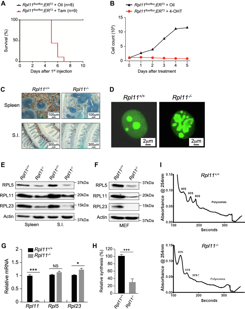
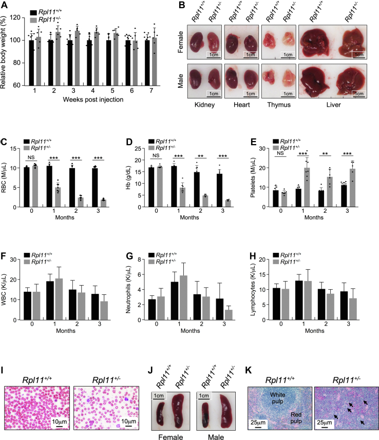
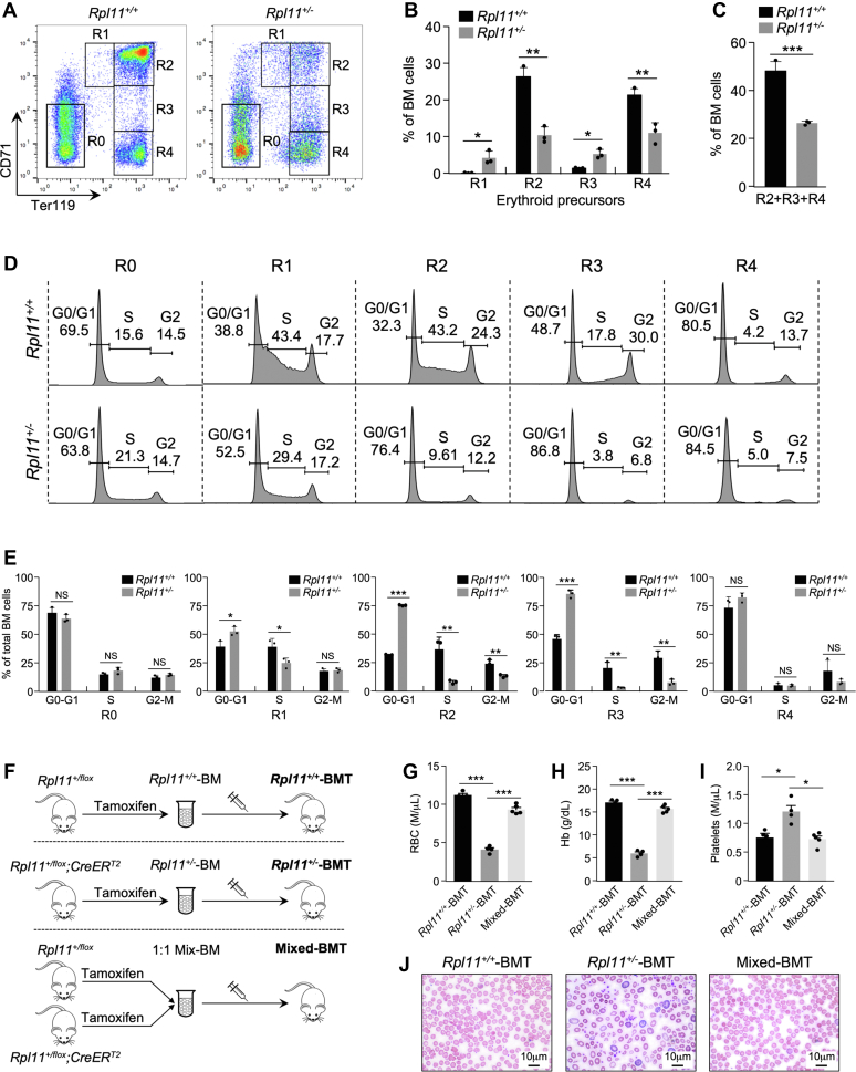
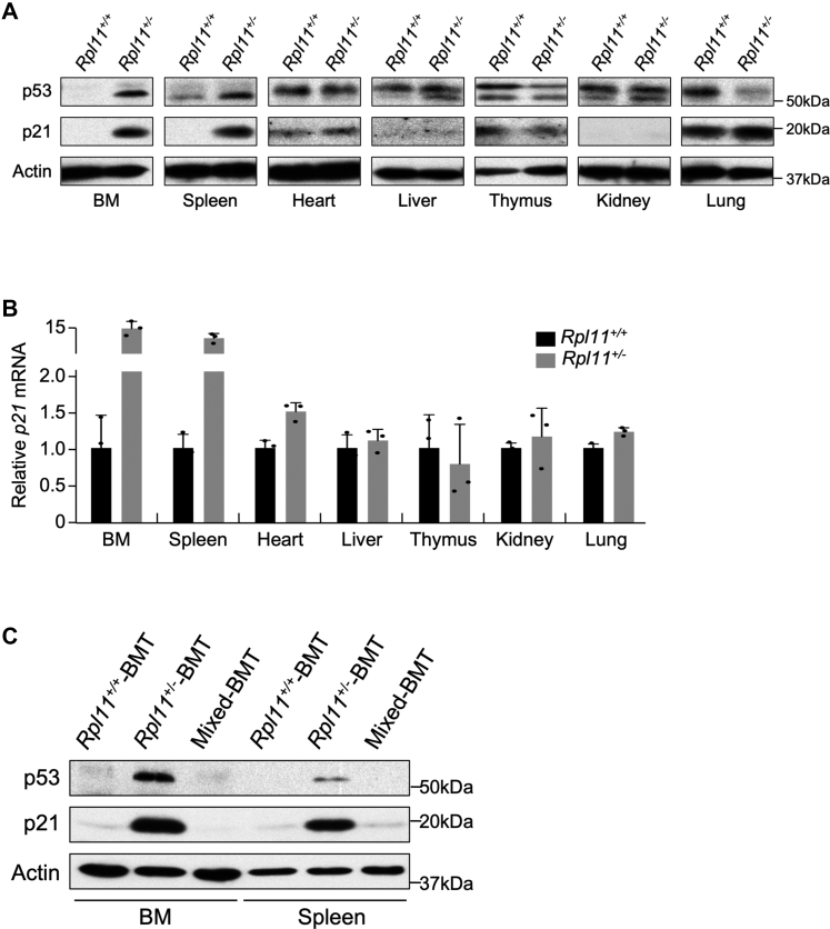
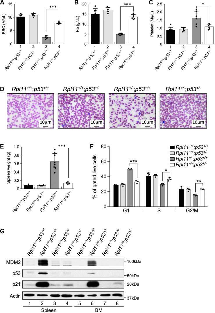
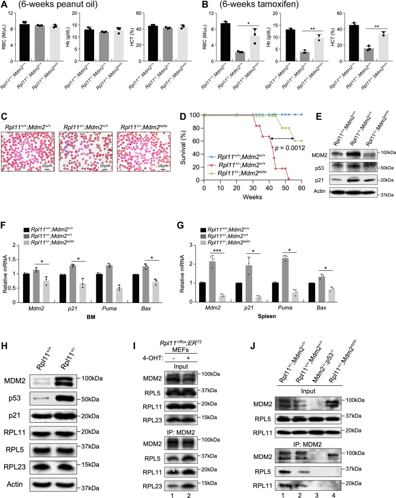
Recent discovery of the ribosomal protein (RP) RPL11 interacting with and inhibiting the E3 ubiquitin ligase function of MDM2 established the RP-MDM2-p53 signaling pathway, which is linked to biological events, including ribosomal biogenesis, nutrient availability, and metabolic homeostasis. Mutations in RPs lead to a diverse array of phenotypes known as ribosomopathies in which the role of p53 is implicated. Here, we generated conditional RPL11-deletion mice to investigate in vivo effects of impaired RP expression and its functional connection with p53. While deletion of one Rpl11 allele in germ cells results in embryonic lethality, deletion of one Rpl11 allele in adult mice does not affect viability but leads to acute anemia. Mechanistically, we found RPL11 haploinsufficiency activates p53 in hematopoietic tissues and impedes erythroid precursor differentiation, resulting in insufficient red blood cell development. We demonstrated that reducing p53 dosage by deleting one p53 allele rescues RPL11 haploinsufficiency-induced inhibition of erythropoietic precursor differentiation and restores normal red blood cell levels in mice. Furthermore, blocking the RP-MDM2-p53 pathway by introducing an RP-binding mutation in MDM2 prevents RPL11 haploinsufficiency-caused p53 activation and rescues the anemia in mice. Together, these findings demonstrate that the RP-MDM2-p53 pathway is a critical checkpoint for RP homeostasis and that p53-dependent cell cycle arrest of erythroid precursors is the molecular basis for the anemia phenotype commonly associated with RP deficiency.

Keywords: MDM2; RPL11; anemia; p53; ribosomal protein; ribosomopathy.


Conflict of interest statement

Conflict of interest The authors declare that they have no conflicts of interest with the contents of this article.

Figures

Figure 1
Figure 1
Homozygous deletion of RPL11 in mice inhibits protein synthesis and causes rapid lethality.A, mice bearing RPL11 homozygous deletion exhibit early lethality. Kaplan–Meier curve depicting the survival of Rpl11flox/flox;CreERT2 mice treated with three injections of peanut oil (n = 8) or tamoxifen (n = 9). B, Rpl11flox/flox;CreERT2 MEF cells were seeded into 10-cm tissue culture dishes at 1 × 105 cells/dish. Peanut oil or 4-hydroxitamoxifen (4-OHT) was added to each dish at day-0. Every day, cells were harvested from one dish and counted using a Bio-Rad cell counter. C, representative Ki-67-stained sections from paraffin-embedded WT and Rpl11−/− mouse tissues isolated after treatment with peanut oil or tamoxifen. The normal architecture of the spleen and small intestine (S.I.) and positive Ki-67 signals were seen in the WT mouse tissues. The architectures of the tissues were destroyed, and the Ki-67 signals were markedly reduced in the Rpl11−/− mouse tissues. D, structure of nucleolus of WT and Rpl11−/− cells. Early passages of WT and Rpl11−/− MEF cells were fixed and immune-stained with B23 (nucleophosmin, NPM) antibody to visualize nucleolar structure. (Original magnification 400×). E, deletion of RPL11 inhibits protein expression. Rpl11flox/flox;CreERT2 mouse spleen and small intestine (S.I.) tissues were harvested at day 5 after the first peanut oil or tamoxifen injection. Protein levels in crude tissue extracts were analyzed by Western blot analysis using antibodies against various proteins as indicated. F, WT and Rpl11−/− MEF cells were analyzed by Western blot using antibodies against various proteins as indicated. G, quantitative real-time RT-PCR analysis for relative levels of mRNA of mouse bone marrow tissues. The tissues were harvested from Rpl11flox/flox;CreERT2 mice at day 5 after the first peanut oil or tamoxifen injection, and total RNA were extracted for analysis. (n = 3 per group; error bars represent standard deviation. NS: nonsignificant; ∗p < 0.05; ∗∗∗p < 0.001; Student’s t test. H, protein synthesis rates in WT and Rpl11−/− MEF cells were assessed by [35S]-methionine incorporation. The number of counts was normalized to total protein content. (n = 3 per group; error bars represent standard deviation) ∗∗∗p < 0.001; Student’s t test. I, polysome profiling of the Rpl11+/+ and Rpl11−/− MEF cells. The cells were lysed, and the nuclei, mitochondria, and large cellular debris were removed via centrifugation. The remaining supernatant was loaded onto a linear sucrose gradient, centrifuged at 32,000 rpm for 2 h to separate ribosomal subunits, monosomes, and polysomes. Ribosome profiles were obtained by measuring the absorbance of RNA at 254 nm.
Figure 2
Figure 2
Heterozygous deletion of RPL11 in adult mice impinges on tissues of hematopoiesis.A, effect of RPL11 heterozygous deletion on body weight of adult mice. Four-month-old male Rpl11+/+;CreERT2 (n = 11) and Rpl11+/flox;CreERT2 (n = 12) mice were given three injections of tamoxifen every other day. Changes in body weight were recorded every week after the injection. Data were presented as ± standard error of the mean. No significant differences were observed. B, images of kidney, heart, thymus, and liver tissues freshly isolated from female and male Rpl11+/+;CreERT2 and Rpl11+/flox;CreERT2 mice 2 months after treatment with tamoxifen. C–H, Four-month-old male Rpl11+/+;CreERT2 and Rpl11+/flox;CreERT2 mice were treated with tamoxifen injection every other day for 3 times. Peripheral blood cells were collected every month after the treatment for 3 months and measured for red blood cell (RBC) count (C), hemoglobin (Hb) content (D), platelet count (E), white blood cell (WBC) count (F), neutrophil count (G), and lymphocyte count (H). Results are show as mean ± standard deviation from a minimum of five mice per group. (n = 3 per group; error bars represent standard deviation; NS, not significant, ∗∗p < 0.01, ∗∗∗p < 0.001; Student’s t test). I, peripheral blood smears from Rpl11+/+;CreERT2 and Rpl11+/flox;CreERT2 mice 2 months after treatment with tamoxifen. Original magnification (1000×). J, images of representative spleens from 4-month-old female and male Rpl11+/+;CreERT2 and Rpl11+/flox;CreERT2 mice 2 months after treatment with tamoxifen. K, hematoxylin and eosin staining of representative spleen sections 2 months after tamoxifen injection. The images showing disruption of the red/white pulp structures in the Rpl11+/− mice compared to Rpl11+/+ mice. Arrows indicate megakaryocytes. Original magnification (200×). Rpl11+/flox, RPL11 heterozygous flox mice.
Figure 3
Figure 3
RPL11 haploinsufficiency impairs erythropoiesis by causing G1 cell cycle arrest in erythroid cells.A, representative CD71/Ter119 flow cytometric profiles of BM cells isolated from Rpl11+/+ and Rpl11+/− mice. Gates of different erythroblast populations were according to expression levels of CD71 and Ter119. R0 contains mixed populations of hematopoietic stem cells and early progenitors. R1, R2, R3, and R4 represent proerythroblasts, basophilic, polychromatic, and orthochromatic erythroblasts, respectively. B, bar graphs representing the percent of total cells of the R1-R4 subpopulations. (n = 3 per group; error bars represent standard deviation; ∗p < 0.05; ∗∗p < 0.01; Student’s t test). C, bar graph representing the percentage of Ter119high (R2+R3+R4) cells from total bone marrow. (n = 3 per group; error bars represent standard deviation, ∗∗∗p < 0.001; Student’s t test). D, BM extracted from Rpl11+/+ and Rpl11+/− mice were incubated with CD71 and Ter119 antibodies followed by cell sorting to separate R0-R4 populations. Flow cytometry was applied to analyze the cell cycle distribution of individual erythroid progenitor populations. Quantification of the cell cycle distribution from each phase was shown as a percent of total cells. E, bar graphs represent the average percentage of cells from erythroid progenitor subsets (R0-R4) in each stage of the cell cycle. (n = 3 per group; error bars represent standard deviation; NS: nonsignificant; ∗p < 0.05; ∗∗p < 0.01; ∗∗∗p < 0.001; Student’s t test). F, schematic representation of the BM transplantation (BMT) procedure. Top: experimental outline of murine BM reconstitution assay with WT (Rpl11+/+) donor mice. Middle: experimental procedure of BM reconstitution assay with heterozygous deletion RPL11 (Rpl11+/−) donor mice. Bottom: experimental procedure of BM reconstitution assay with donor BM isolated from WT and heterozygous deletion RPL11 mice mixed at 1:1 ratio. All recipient mice were treated with 8-Gy γ-irradiation prior to transplantation. G–I, peripheral blood analysis of mice after BMT. Peripheral blood was extracted from BMT-mice 2 months post transplantation for total blood count analysis of RBC (G), hemoglobin (Hb) (H), and platelet (I) from Rpl11+/+-BMT (black bar), Rpl11+/−-BMT (gray bar), and Mixed-BMT (white bar) mice. Results are shown as mean ± standard deviation from a minimum of three mice per group. J, peripheral blood smears from Rpl11+/+-BMT, Rpl11+/−-BMT, and Mixed-BMT mice 2 months after transplantation. Original magnification (1000×). BM, bone marrow.
Figure 4
Figure 4
RPL11 haploinsufficiency activates p53 in hematopoietic tissues.A, tissue expression pattern of p53 and p21 proteins in Rpl11+/+ and Rpl11+/− mice. Tissues were isolated from adult Rpl11+/+;CreERT2 and Rpl11+/flox;CreERT2 mice after tamoxifen treatment for 2 weeks prior to protein extraction for immunoblot analysis of p53, p21, and actin. B, Rpl11+/+ and Rpl11+/− mice were treated, and tissues were isolated as in (A). Tissue expression pattern of p21 mRNA in Rpl11+/+ and Rpl11+/− mice was analyzed by RT-PCR. C, Bone marrow (BM) and spleen tissues were harvested from BM transplant mice and proteins were extracted for immunoblot analysis of MDM2, p53, p21, and actin. Rpl11+/flox, RPL11 heterozygous flox mice.
Figure 5
Figure 5
Deletion of one p53 allele rescues RPL11 haploinsufficiency–induced anemia and restores cell cycle progression in erythroid cells.A–C, Six-week old Rpl11+/+;CreERT2;p53+/+, Rpl11+/flox;CreERT2;p53+/+, Rpl11+/+;CreERT2;p53+/−, and Rpl11+/flox;CreERT2;p53+/− male mice were injected with tamoxifen every other day for 3 times to generate Rpl11+/+;p53+/+, Rpl11+/+;p53+/−, Rpl11+/−;p53+/+, and Rpl11+/−;p53+/− mice. Peripheral blood was isolated 2 months after the tamoxifen treatment and analyzed for RBC (A), hemoglobin (Hb) (B), and platelet (C). (n = 3 per group; error bars represent standard deviation; ∗p < 0.05; ∗∗∗p < 0.001; Student’s t test). D, peripheral blood was isolated from abovementioned mice, and blood smears were performed. Original magnification (1000×). E, spleen weight in grams from abovementioned mice. (n = 3 per group; error bars represent standard deviation; ∗∗∗p < 0.001; Student’s t test). F, BM cells were extracted from abovementioned mice and gated for erythroid precursors. The gated cells were analyzed for cell cycle distribution. (n = 3 per group; error bars represent standard deviation; ∗p < 0.05; ∗∗p < 0.01; ∗∗∗p < 0.001; Student’s t test). G, BM and spleen tissues were harvested from abovementioned mice, and tissue extracts were analyzed by Western blot for MDM2, p53, p21 and actin. BM, bone marrow; Rpl11+/flox, RPL11 heterozygous flox mice.
Figure 6
Figure 6
RPL11 haploinsufficiency–induced anemia involves the RP-MDM2-p53 pathway.A–B, Four-month-old Rpl11+/+;CreERT2;Mdm2+/+, Rpl11+/flox;CreERT2;Mdm2+/+, and Rpl11+/flox;CreERT2;Mdm2m/m male mice were treated with peanut oil (A) or tamoxifen (B) for 6 weeks. Peripheral blood was isolated and analyzed for RBC, hemoglobin (Hb), and hematocrit (HCT). (n = 3 per group; error bars represent standard deviation; ∗p < 0.05; ∗∗p < 0.01; Student’s t test). C, peripheral blood was isolated from abovementioned mice, and blood smears were performed. Original magnification (1000×). D, Kaplan–Meier curve depicting the survival of Rpl11+/+;CreERT2;Mdm2+/+ (n = x), Rpl11+/flox;CreERT2;Mdm2+/+ (n = x), and Rpl11+/flox;CreERT2;Mdm2m/m (n = x), male mice treated with three injections of tamoxifen. E, BM tissues were harvested from 4-month-old Rpl11+/+;CreERT2;Mdm2+/+, Rpl11+/flox;CreERT2;Mdm2+/+, and Rpl11+/flox;CreERT2;Mdm2m/m male mice 2 weeks after treated with three injections of tamoxifen. Tissue extracts were analyzed by Western blot for protein expression of MDM2, p53, p21, and actin. F–G, quantitative real-time RT-PCR analyses of Mdm2, p21, Puma, and Bax transcript levels in BM (F) and Spleen (G) tissues isolated from 4-month-old Rpl11+/+;CreERT2;Mdm2+/+, Rpl11+/flox;CreERT2;Mdm2+/+, and Rpl11+/flox;CreERT2;Mdm2m/m male mice 2 weeks after treated with three injections of tamoxifen. GAPDH transcripts were used for normalization. (n = 3 per group; error bars represent standard deviation; ∗p < 0.05; ∗∗∗p < 0.001; Student’s t test). H, WT and Rpl11+/− MEF cells were analyzed by Western blot using antibodies against various proteins as indicated. I, Rpl11+/flox;ERT2 MEFs were exposed to 4-OHT for 20 h prior to immunoprecipitation with anti- MDM2 antibodies and immunoblotting for MDM2, RPL5, RPL11, and RPL23. J, BM tissues were isolated from Rpl11+/+;Mdm2+/+, Rpl11+/−;Mdm2+/+, Rpl11+/+;Mdm2−/−;p53−/−, and Rpl11+/−;Mdm2m/m mice; MDM2 immunoprecipitation was carried out prior to immunoblot analysis for MDM2, RPL5, and RPL11. BM, bone marrow; Rpl11+/flox, RPL11 heterozygous flox mice.

Similar articles

Cited by

References

    1. Hollstein M., Sidransky D., Vogelstein B., Harris C.C. p53 mutations in human cancers. Science. 1991;253:49–53. - PubMed
    1. Olivier M., Hollstein M., Hainaut P. TP53 mutations in human cancers: origins, consequences, and clinical use. Cold Spring Harbor Perspect. Biol. 2010;2:a001008. - PMC - PubMed
    1. Lane D.P. p53, guardian of the genome. Nature. 1992;358:15–16. - PubMed
    1. Moll U.M., Petrenko O. The MDM2-p53 interaction. Mol. Cancer Res. 2003;1:1001–1008. - PubMed
    1. Zhang Y., Lu H. Signaling to p53: ribosomal proteins find their way. Cancer Cell. 2009;16:369–377. - PMC - PubMed

Publication types

Substances