. 2022 Nov 26;13(11):1002.
doi: 10.1038/s41419-022-05449-6.

Mutual regulation between phosphofructokinase 1 platelet isoform and VEGF promotes glioblastoma tumor growth

Affiliations

Mutual regulation between phosphofructokinase 1 platelet isoform and VEGF promotes glioblastoma tumor growth

Je Sun Lim et al. Cell Death Dis. .

Abstract

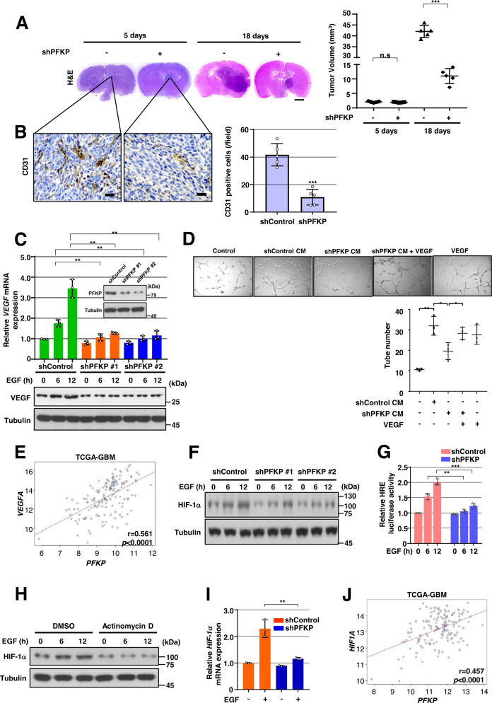
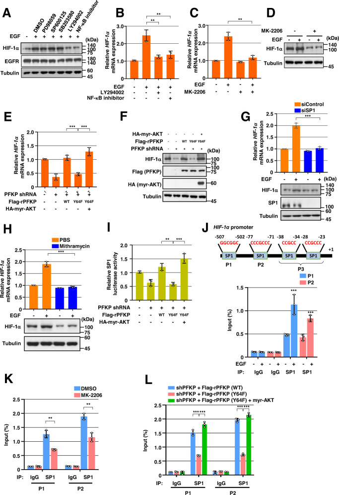
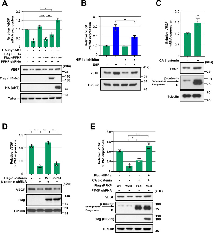
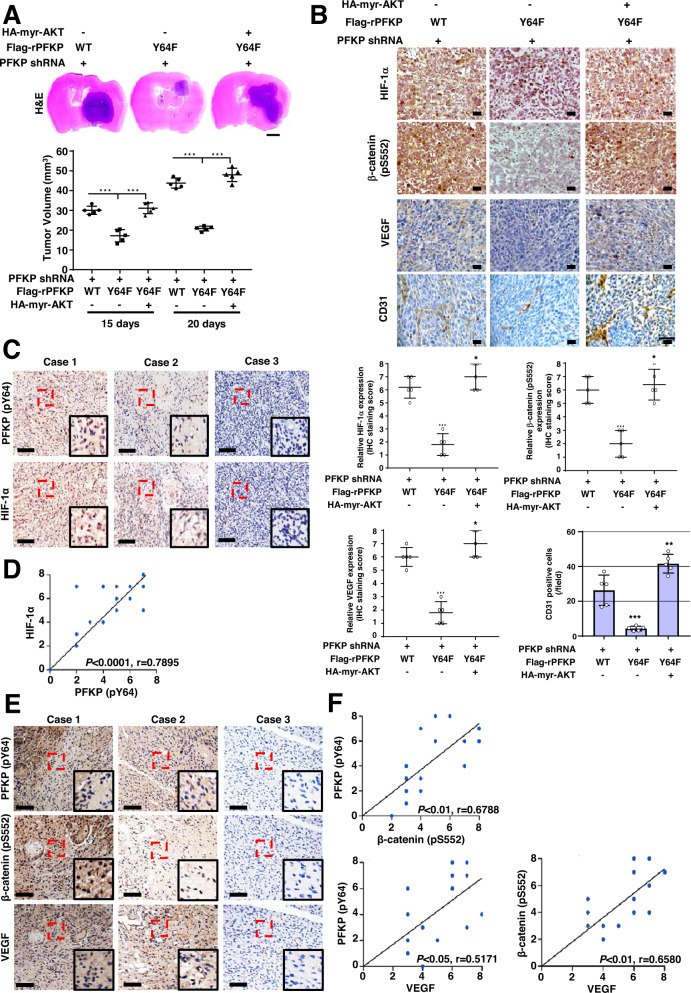
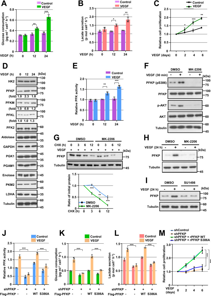
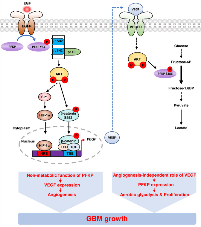
Glioblastoma (GBM) is a highly vascular malignant brain tumor that overexpresses vascular endothelial growth factor (VEGF) and phosphofructokinase 1 platelet isoform (PFKP), which catalyzes a rate-limiting reaction in glycolysis. However, whether PFKP and VEGF are reciprocally regulated during GBM tumor growth remains unknown. Here, we show that PFKP can promote EGFR activation-induced VEGF expression in HIF-1α-dependent and -independent manners in GBM cells. Importantly, we demonstrate that EGFR-phosphorylated PFKP Y64 has critical roles in both AKT/SP1-mediated transcriptional expression of HIF-1α and in the AKT-mediated β-catenin S552 phosphorylation, to fully enhance VEGF transcription, subsequently promoting blood vessel formation and brain tumor growth. Levels of PFKP Y64 phosphorylation in human GBM specimens are positively correlated with HIF-1α expression, β-catenin S552 phosphorylation, and VEGF expression. Conversely, VEGF upregulates PFKP expression in a PFKP S386 phosphorylation-dependent manner, leading to increased PFK enzyme activity, aerobic glycolysis, and proliferation in GBM cells. These findings highlight a novel mechanism underlying the mutual regulation that occurs between PFKP and VEGF for promoting GBM tumor growth and also suggest that targeting the PFKP/VEGF regulatory loop might show therapeutic potential for treating GBM patients.


Conflict of interest statement

The authors declare no competing interests.

Figures

Fig. 1
Fig. 1. PFKP expression is required for EGFR activation-induced VEGF expression in vitro and GBM angiogenesis in vivo.
WB and qRT-PCR were performed with indicated primers and antibodies, respectively (C, F, H, I). A, B Representative H&E staining images of intracranial xenografts bearing LN229/EGFRvIII cells stably expressing with or without PFKP shRNA (A; left panel) and quantification of tumor volumes (A; right panel). IHC analyses of the tumor tissues with an anti-CD31 antibody (B; left panel). Quantification of CD31 (B; right panel). Scale bar, 2 mm (A) and 100 μm (B). C GSCs with different shRNAs against PFKP (inside panel). Serum-starved GSCs with or without PFKP depletion were treated with EGF (100 ng/mL). D HUVECs were treated with or without a conditioned medium (CM) from control shRNA-expressing or PFKP shRNA-expressing LN229/EGFRvIII cells in the presence or absence of VEGF (20 ng/mL). HUVECs tube formation was observed. Representative images were acquired under an optical microscope (50×), and the tube number (/field) was quantified. E TCGA analysis of PFKP and VEGF mRNA expression from TCGA-GBM data (n = 154). F Serum-starved GSCs with or without PFKP depletion were treated with EGF (100 ng/mL). G HRE luciferase activity in GSCs with stable expression of control shRNA or PFKP shRNA was measured. H Serum-starved GSCs were pretreated with DMSO or actinomycin D (1 μg/mL) for 1 h and then stimulated with or without EGF (100 ng/mL). I Serum-starved GSCs stably expressing control shRNA, or PFKP shRNA were treated with or without EGF (100 ng/mL) for 12 h. J TCGA analysis of PFKP and HIF-1α mRNA expression from TCGA-GBM data (n = 154). Data are presented as mean ± standard deviation of three independent experiments (A, C, D, G, I). ***P < 0.001, based on the Student’s t-test.
Fig. 2
Fig. 2. PFKP Y64 phosphorylation induces EGFR activation-enhanced HIF-1α transcriptional expression through SP1 transactivation.
WB and qRT-PCR were performed with indicated primers and antibodies, respectively (AH). A Serum-starved GSCs were pretreated DMSO, PD98059 (20 μM), SP600125 (25 μM), SB203580 (10 μM), LY294002 (20 μM), or NF-κB inhibitor (1 μM) for 1 h and then stimulated with or without EGF (100 ng/mL) for 12 h. BD Serum-starved GSCs were pretreated DMSO, LY294002, NF-κB inhibitor (B), or MK-2206 (5 μM) (C, D) for 1 h and then stimulated with or without EGF (100 ng/mL) for 12 h. E, F LN229/EGFRvIII cells with or without PFKP depletion and with or without reconstituted expression of WT Flag-rPFKP or Flag-rPFKP Y64F mutant in the presence or absence of HA-myr-AKT expression. G GSCs were transfected with control siRNA or SP1 siRNA. H GSCs were treated with PBS or mithramycin (500 nM) for 12 h. I SP1 luciferase activity in LN229/EGFRvIII cells with or without PFKP depletion and with or without reconstituted expression of WT Flag-rPFKP or Flag-PFKP Y64F mutant in the presence or absence of HA-myr-AKT expression was measured. JL ChIP assays were performed with anti-SP1 antibody, and real-time PCR analyses were performed with primers against the HIF-1α promoter. (J) A schematic of the putative SP1 binding site (Marked as P1–P3) on the HIF-1α promoter region (J; upper panel). GSCs were treated with or without EGF (100 ng/mL) for 12 h (J; bottom panel). K LN229/EGFRvIII cells were pretreated with DMOS or MK-2206 (5 μM) for 1 h and then treated with EGF (100 ng/ml) for 12 h. L LN229/EGFRvIII cells with PFKP depletion and with or without reconstituted expression of WT Flag-rPFKP or Flag-rPFKP Y64F mutant were transfected with or without HA-myr-AKT. Data are presented as mean ± standard deviation of three independent experiments (B, C, E, G, HL). ***P < 0.001, based on the Student’s t-test or one-way ANOVA with Tukey’s post hoc test.
Fig. 3
Fig. 3. PFKP Y64 phosphorylation induces VEGF expression through HIF-1 α expression and β-catenin Ser552 phosphorylation in response to EGFR activation.
WB and qRT-PCR were performed with indicated primers and antibodies, respectively (AE). A LN229/EGFRvIII cells with or without PFKP depletion and with or without reconstituted expression of WT Flag-rPFKP or Flag-rPFKP Y64F mutant were transfected with or without Flag-HIF-1α and HA-myr-AKT. B Serum starved LN229 cells were pretreated with DMSO or HIF inhibitor (10 μM) for 1 h and then treated with EGF (100 ng/mL) for 12 h. C LN229 cells were transfected with a control vector or CA β-catenin. D LN229/EGFRvIII cells with stable expression of β-catenin shRNA or a control shRNA were reconstituted with or without WT rβ-catenin or rβ-catenin S552A mutant. E PFKP-depleted LN229/EGFRvIII cells with or without reconstituted expression of WT Flag-rPFKP or Flag-rPFKP Y64F mutant were transfected with or without CA β-catenin and Flag- HIF-1α. Data are presented as mean ± standard deviation of three independent experiments (AE). **P < 0.01; ***P < 0.001, based on the Student’s t-test or one-way ANOVA with Tukey’s post hoc test.
Fig. 4
Fig. 4. PFKP Y64 phosphorylation induces HIF-1 α expression, β-catenin S552 phosphorylation, and VEGF expression and promotes blood vessel formation in vivo.
A Representative H&E staining images of intracranial xenografts bearing PFKP-depleted LN229/EGFRvIII cells with or without reconstituted expression of WT Flag-rPFKP or Flag-rPFKP Y64F mutant with or without myr-AKT1 (A; upper panel) and quantification of tumor volumes (A; bottom panel). Scale bar, 2 mm. B IHC analyses of tumor tissues with the indicated antibodies (upper panel). Quantification of indicated IHC staining (bottom panel). Scale bar, 100 μm. C, E IHC staining of human GBM specimens was performed with indicated antibodies (n = 25). Representative images from the staining of three different specimens are shown. Scale bar, 100 μm. D, F The IHC stains were scored, and correlation analyses were performed. The Pearson correlation test was used. Note that the scores of some samples overlapped. ***P < 0.001, based on one-way ANOVA with Tukey’s post hoc test.
Fig. 5
Fig. 5. VEGF induces PFKP expression, PFK enzyme activity, aerobic glycolysis, and proliferation in GBM cells.
WB and qRT-PCR were performed with indicated primers and antibodies, respectively (D, FI). A, B Serum-starved GSCs were treated with VEGF (20 ng/mL). Glucose consumption (A) and lactate secretion (B) were analyzed. C GSCs in 0.1% serum medium were treated with VEGF (20 ng/mL). WST-8 assay was then performed. D, E Serum-starved GSCs were treated with VEGF (20 ng/mL). Indicated protein expression levels (D) and PFK enzymatic activity (E) were measured. F Serum-starved GSCs were pretreated with DMSO or MK-2206 (5 μM) for 1 h and then stimulated with VEGF (20 ng/mL) for 30 min. G Serum-starved GSCs were pretreated with VEGF (20 ng/mL) for 1 h and then treated with cycloheximide (CHX;100 μg/mL) in the presence of DMSO or MK-2206 (5 μM). Quantification of PFKP levels relative to tubulin is shown (bottom panel). H Serum-starved GSCs were pretreated with DMSO or MK-2206 (5 μM) for 1 h and then stimulated with or without VEGF (20 ng/mL) for 24 h. I Serum-starved GSCs were pretreated with DMSO or SU1498 (30 μM) for 1 h and then stimulated with or without VEGF (20 ng/mL) for 24 h. JL LN229 cells with or without expression of PFKP shRNA and with or without the reconstituted expression of WT Flag-rPFKP or Flag-rPFKP S386A were cultured in serum-free DMEM with or without VEGF (20 ng/mL) for 24 h. PFK enzymatic activity (J), glucose consumption (K), and lactate secretion (L) were then analyzed. M LN229 cells with or without the expression of PFKP shRNA and with or without the reconstituted expression of WT Flag-rPFKP or Flag-rPFKP S386A were cultured in 0.1% serum medium with or without VEGF (20 ng/mL). WST-8 assay was then performed. Data are presented as mean ± SD of three independent experiments (AC, E, G, JM). *P < 0.05; **P < 0.01; ***P < 0.001, based on the Student’s t-test or one-way ANOVA with Tukey’s post hoc test.
Fig. 6
Fig. 6. Reciprocal regulation between PFKP and VEGF promotes GBM tumor growth.
A schematic of the proposed reciprocal regulation that occurs between PFKP and VEGF for regulating GBM tumor growth. HRE hypoxia response element, TBE TCF binding element.

References

    1. Apte RS, Chen DS, Ferrara N. VEGF in signaling and disease: beyond discovery and development. Cell. 2019;176:1248–64. doi: 10.1016/j.cell.2019.01.021. - DOI - PMC - PubMed
    1. Hanahan D, Weinberg RA. Hallmarks of cancer: the next generation. Cell. 2011;144:646–74. doi: 10.1016/j.cell.2011.02.013. - DOI - PubMed
    1. Jain RK, di Tomaso E, Duda DG, Loeffler JS, Sorensen AG, Batchelor TT. Angiogenesis in brain tumours. Nat Rev Neurosci. 2007;8:610–22. doi: 10.1038/nrn2175. - DOI - PubMed
    1. Pouyssegur J, Dayan F, Mazure NM. Hypoxia signalling in cancer and approaches to enforce tumour regression. Nature. 2006;441:437–43. doi: 10.1038/nature04871. - DOI - PubMed
    1. Shweiki D, Itin A, Soffer D, Keshet E. Vascular endothelial growth factor induced by hypoxia may mediate hypoxia-initiated angiogenesis. Nature. 1992;359:843–5. doi: 10.1038/359843a0. - DOI - PubMed

Publication types

MeSH terms