

VEP examination with new portable device
- PMID: 36436114
- PMCID: PMC9911502
- DOI: 10.1007/s10633-022-09911-w
VEP examination with new portable device
Abstract
Introduction: We developed a new portable device called "VEPpeak" for the examination of visual evoked potentials (VEPs) to extend VEP examination beyond specialized electrophysiological laboratories and to simplify the use of this objective, noninvasive, and low-cost method for diagnostics of visual and central nervous system dysfunctions.
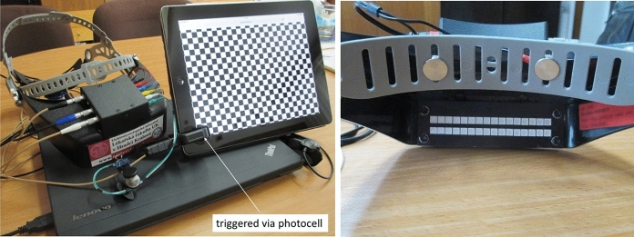
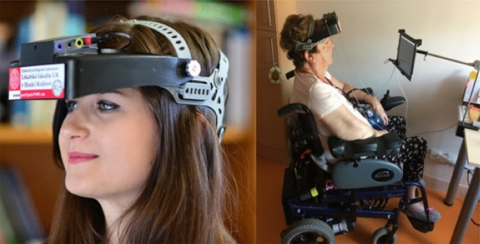
Methods: VEPpeak consists of a plastic headset with a total weight of 390 g containing four EEG amplifiers, an A/D converter, a control unit, and a visual LED stimulator built in the front, vertically adjustable peak. The device is powered and controlled via USB connection from a standard PC/notebook using custom software for visual stimuli generation and for VEP recording and processing. Up to four electrodes can be placed at any scalp location or in combination with two dry electrodes incorporated into the headset. External visual stimulators, such as a tablet, can be used with synchronization. Feasibility and validation studies were conducted with 86 healthy subjects and 76 neuro-ophthalmological patients including 67 who were during the same session also tested with a conventional VEP system.
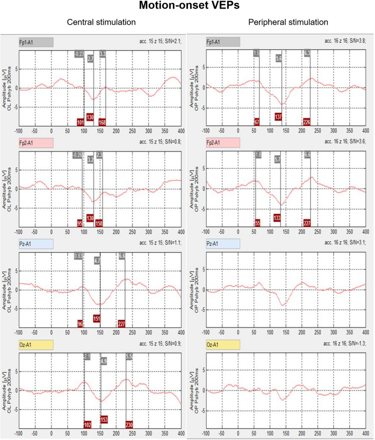
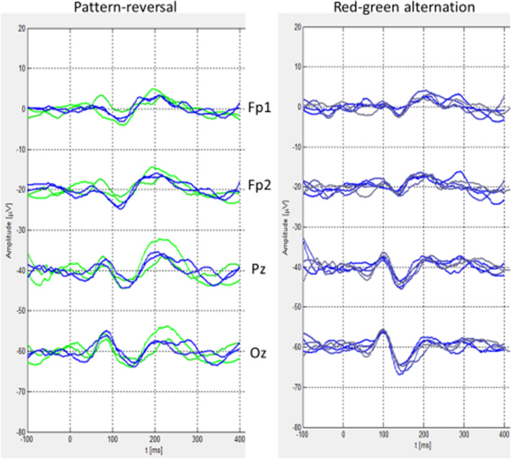
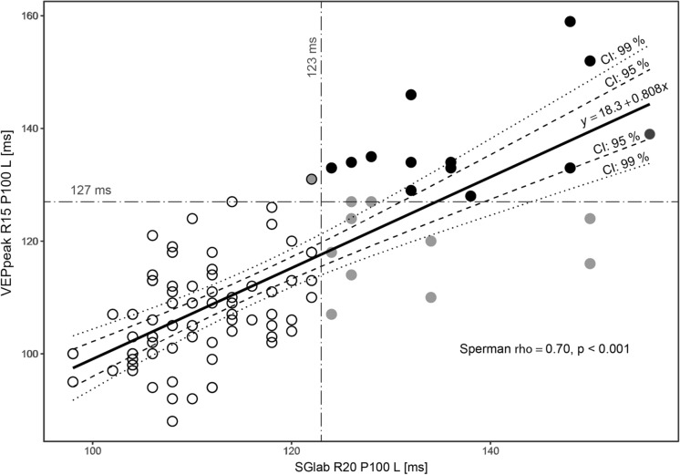
Results: VEPpeak recordings to standard (pattern-reversal) and non-standard (motion-onset, red-green alternation) were robust and repeatable and obtained also in immobilized patients. Good comparability of results was achieved between VEPpeak and standard examination. Some systematic differences in peak latencies and amplitudes are consistent with differences in stimulus characteristics of the two compared systems.
Discussion: VEPpeak provides an inexpensive system for clinical use requiring portability. In addition to ISCEV standard VEP protocols, free choice of stimuli and bio-signal recordings make the device universal for many electrophysiological purposes.
Keywords: Cognitive ERP; Motion-onset; Pattern-reversal; VEP diagnostics; VEP portable device; VEPpeak.
© 2022. The Author(s).
Conflict of interest statement
All authors certify that they have no affiliations with or involvement in any organization or entity with any financial interest (such as honoraria; educational grants; participation in speakers’ bureaus; membership, employment, consultancies, stock ownership, or other equity interest; and expert testimony or patent-licensing arrangements), or nonfinancial interest (such as personal or professional relationships, affiliations, knowledge, or beliefs) in the subject matter or materials discussed in this manuscript.
Figures

Similar articles
-
New portable device for an examination of visual cognitive evoked potentials might extend their diagnostic applications in psychiatry.Psychiatry Res Neuroimaging. 2024 Jan;337:111768. doi: 10.1016/j.pscychresns.2023.111768. Epub 2023 Dec 15. Psychiatry Res Neuroimaging. 2024. PMID: 38128365
-
Evaluation of Diurnal Changes of Mental Fatigue Using a New Portable Device for Visual Cognitive Evoked Potentials.Acta Medica (Hradec Kralove). 2023;66(2):55-60. doi: 10.14712/18059694.2023.16. Acta Medica (Hradec Kralove). 2023. PMID: 37930094
-
Improving reproducibility of VEP recording in rats: electrodes, stimulus source and peak analysis.Doc Ophthalmol. 2011 Oct;123(2):109-19. doi: 10.1007/s10633-011-9288-8. Epub 2011 Sep 10. Doc Ophthalmol. 2011. PMID: 21909708
-
Visually evoked potentials.Handb Clin Neurol. 2019;160:501-522. doi: 10.1016/B978-0-444-64032-1.00034-5. Handb Clin Neurol. 2019. PMID: 31277872 Review.
-
Visually evoked potentials and electroretinography in neurologic evaluation.Neurol Clin. 1991 Feb;9(1):225-42. Neurol Clin. 1991. PMID: 1849226 Review.
Cited by
-
Can a Portable Flash Visual Evoked Potential (VEP) Device Identify Chiasmal Decussation Anomalies in Albinism?Diagnostics (Basel). 2025 May 30;15(11):1395. doi: 10.3390/diagnostics15111395. Diagnostics (Basel). 2025. PMID: 40506967 Free PMC article.
-
Visual Evoked Potentials for the Detection of Diabetic Retinal Neuropathy.Int J Mol Sci. 2023 Apr 17;24(8):7361. doi: 10.3390/ijms24087361. Int J Mol Sci. 2023. PMID: 37108524 Free PMC article. Review.
References
Publication types
MeSH terms
LinkOut - more resources
Full Text Sources
