Review
. 2022 Nov 27;79(12):607.
doi: 10.1007/s00018-022-04630-6.

Mechanisms of colorectal liver metastasis development

Affiliations
Review

Mechanisms of colorectal liver metastasis development

Tal Shasha et al. Cell Mol Life Sci. .

Abstract

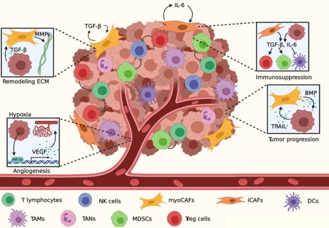
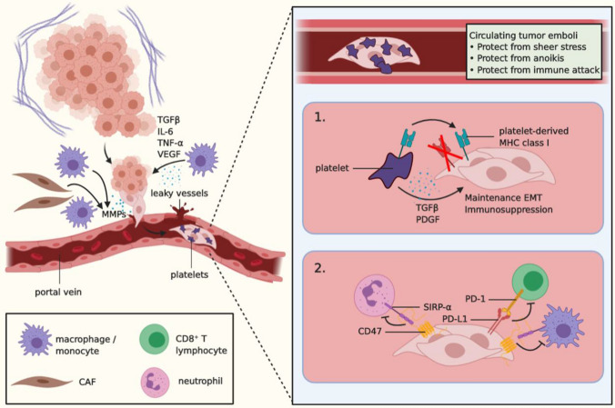
Colorectal cancer (CRC) is a leading cause of cancer-related death worldwide, largely due to the development of colorectal liver metastases (CRLM). For the establishment of CRLM, CRC cells must remodel their tumor-microenvironment (TME), avoid the immune system, invade the underlying stroma, survive the hostile environment of the circulation, extravasate into the liver, reprogram the hepatic microenvironment into a permissive pre-metastatic niche, and finally, awake from a dormant state to grow out into clinically detectable CRLM. These steps form part of the invasion-metastasis cascade that relies on reciprocal interactions between the tumor and its ever-changing microenvironment. Such interplay provides a strong rational for therapeutically targeting the TME. In fact, several TME constituents, such as VEGF, TGF-β coreceptor endoglin, and CXCR4, are already targeted in clinical trials. It is, however, of utmost importance to fully understand the complex interactions in the invasion-metastasis cascade to identify novel potential therapeutic targets and prevent the establishment of CRLM, which may ultimately greatly improve patient outcome.

Keywords: Cancer; Circulating tumor cells; Epithelial-mesenchymal transition; Invasion-metastasis cascade; Pre-metastatic niche; Tumor microenvironment.


Conflict of interest statement

On behalf of all authors, the corresponding author states that there is no conflict of interest.

Figures

Fig. 1
Fig. 1
Initiation of malignant transformation. As the tumor grows, hypoxia promotes HIF-1α-dependent VEGF transcription and subsequent angiogenesis. TGF-β secreted by tumor cells and fibroblasts may promote differentiation into myoCAFs, degradation of the ECM through MMPs, and immunosuppression by inducing polarization of Treg cells, M2 macrophages, N2 neutrophils, and MDSCs, and inhibition of NK cells and CTLs. SMAD4 CRC cells may express TRAIL, which triggers CAFs to secrete BMP. In turn, BMP signals through Rho and ROCK to further tumor progression. NK cells natural killer cells, CAFs cancer-associated fibroblasts, DCs dendritic cells, TAMs tumor-associated macrophages, TANs tumor-associated neutrophils, MDSCs myeloid-derived suppressor cells, Treg cells regulatory T cells, MMPs matrix metalloproteases, ECM extracellular matrix, VEGF vascular endothelial growth factor, BMP bone morphogenic protein
Fig. 2
Fig. 2
Survival in the circulation. Intravasation of tumor cells into the portal vein is facilitated by TAM-derived signals, such as TGFβ, IL-6, and TNF-α. In addition, TAM-derived VEGF promotes angiogenesis and thus the formation of dysfunctional and leaky vessels, thereby creating a physical entrance for the cancer cells into the circulation. MMPs produced by CAFs, TAMs, and tumor cells can degrade the endothelial basal lamina and cell–cell junctions, which further contributes to intravasation. In the circulation, cancer cells may cluster and form circulating tumor emboli (CTE), which attract platelets and physically shield the cells from sheer stress, anoikis, and immune attack. Furthermore, platelets may produce TGFβ, which promotes maintenance of a more mesenchymal state as well as immunosuppression (1). In addition, platelets may transfer their functional MHC class I complex to tumor cells to further circumvent an immune attack (1). Finally, tumor cells in the circulation may upregulate PD-L1 and CD47, which bind to PD-1 on CTLs, and to SIRP-α on neutrophils and monocytes, respectively, and inhibit these cells from exerting their anti-tumor effects (2). VEGF vascular endothelial growth factor, CAF cancer-associated fibroblast, MMPs matrix metalloproteases, EMT epithelial-mesenchymal transition, MHC major histocompatibility complex, PDGF platelet-derived growth factor, SIRP-α signal-regulatory protein-α, PD-1 programmed cell death protein 1, PD-L1 programmed death-ligand 1
Fig. 3
Fig. 3
CRLM promoting interactions in the liver. A CRC cells navigate through the portal vein and arrive at the fenestrated liver microvasculature, where they extravasate into the liver. Initially, KCs phagocytose the tumor cells. Subsequently, KCs secrete TNFα, which binds to TNFR1 on LSECs and promotes the expression of E-selectin, VCAM, and ICAM-1, facilitating extravasation. Fenestration and leakiness of the vessels can be enhanced by tumor-derived exosomes carrying miR-25-3p, which inhibits KLF4 and KLF2, resulting in dissociation of cell–cell junctions and upregulation of VEGFR2. In addition, neutrophils secrete NETs to capture CTCs in the sinusoids. B Tumor-derived exosomes carrying integrin αvβ5 specifically bind to KCs in the liver, resulting in their secretion of S100P and S100A8, which in turn recruit MDSCs to the hepatic pre-metastatic niche. In addition, MIF-bearing tumor-derived exosomes are taken up by KCs, and stimulate TGF-β expression. This TGF-β activates HSCs and promotes the production of fibronectin, which attracts neutrophils and BMDCs. Presence of TIMP-1 results in increased SDF1/CXCL12 levels, which promote CXCR4-dependent neutrophil recruitment. Furthermore, IL-6-induces JAK/STAT3 signaling in hepatocytes. These release SAA1 and SAA2, which in turn recruit MDSCs. Recruitment of these cells suppresses the hepatic pre-metastatic niche. For example, TGF-β secreted by MDSCs and TAMs, as well as neutrophils and BMDCs, inhibit CD8+ CTL function. In addition, tumor cells downregulate MHC class I and upregulate NK cell decoy molecules to evade immune surveillance. Lactic acid released by tumor cells can decrease intracellular NK cell pH, resulting in apoptosis of and thus evasion from these cells. C Dormant cells exhibit high p38 MAPK and low ERK levels, as well as reduced fibronectin in the ECM and elevated signaling through IFN-γ, FBX8 and SPDEF. CRC cells that awaken and grow out into overt metastases colonizing the liver exhibit low p38 MAPK and high ERK levels, as well as high fibronectin in the ECM and decreased IFN-γ signaling. CRC cells activate liver-specific transcription programs and metabolically adapt to the liver. TGF-β-dependent IL-11 production by CAFs inhibits apoptosis, and supports outgrowth through activation of GP130/STAT3 signaling. LSECs liver sinusoidal endothelial cells, TNFα tumor necrosis factor α, TNFR1 tumor necrosis factor receptor 1, ICAM-1 intracellular adhesion molecule 1, VCAM-1 vascular cell adhesion molecule 1, KLF Krüppel-like Factor, MIF macrophage migration inhibitory factor, MDSC myeloid-derived suppressor cell, BMDC bone marrow-derived cell, TIMP-1 TIMP metallopeptidase inhibitor 1, SAA serum amyloid A, HSC hepatic stellate cell, IFN-γ interferon-γ, FBX8 F-box only protein 8, SPDEF SAM Pointed Domain Containing ETS Transcription Factor

References

    1. Sung H, et al. Global cancer statistics 2020: GLOBOCAN estimates of incidence and mortality worldwide for 36 cancers in 185 countries. CA Cancer J Clin. 2021;71:209–249. doi: 10.3322/caac.21660. - DOI - PubMed
    1. Mehlen P, Puisieux A. Metastasis: a question of life or death. Nat Rev Cancer. 2006;6:449–458. doi: 10.1038/nrc1886. - DOI - PubMed
    1. Manfredi S, et al. Epidemiology and management of liver metastases from colorectal cancer. Ann Surg. 2006;244:254–259. doi: 10.1097/01.sla.0000217629.94941.cf. - DOI - PMC - PubMed
    1. Van Gestel YRBM, et al. Patterns of metachronous metastases after curative treatment of colorectal cancer. Cancer Epidemiol. 2014;38:448–454. doi: 10.1016/j.canep.2014.04.004. - DOI - PubMed
    1. Lemke J, et al. Survival and prognostic factors of colorectal liver metastases after surgical and nonsurgical treatment. Clin Colorectal Cancer. 2016;15:e183–e192. doi: 10.1016/j.clcc.2016.04.007. - DOI - PubMed

Substances