

Utilization of the Pedicled and Free Fibula Flap for Ankle Arthrodesis
- PMID: 36438462
- PMCID: PMC9681623
- DOI: 10.1097/GOX.0000000000004670
Utilization of the Pedicled and Free Fibula Flap for Ankle Arthrodesis
Abstract
Ankle arthrodesis has become a common surgical procedure for individuals with end-stage ankle arthritis, chronic infection, and bony misalignment. Although arthrodesis is typically managed with arthrodesis in situ or realignment, reconstruction may be utilized for patients with more complicated cases that involve metatarsal defects. Our institution utilizes both the pedicled and free fibula flaps for surgical management pertaining to ankle arthrodesis. Our study looks to evaluate the work of a single plastic surgeon and identify patient postoperative outcomes.
Methods: A retrospective chart review was conducted at Beaumont Health System, Royal Oak, for patients who underwent ankle arthrodesis with a pedicled fibula flap for nonunion or avascular necrosis of the talus between the years 2014 and 2022. Demographic data, operative details, complications, medical comorbidities, and patient outcomes were retrospectively gathered and analyzed.
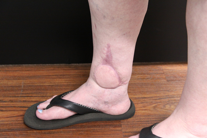
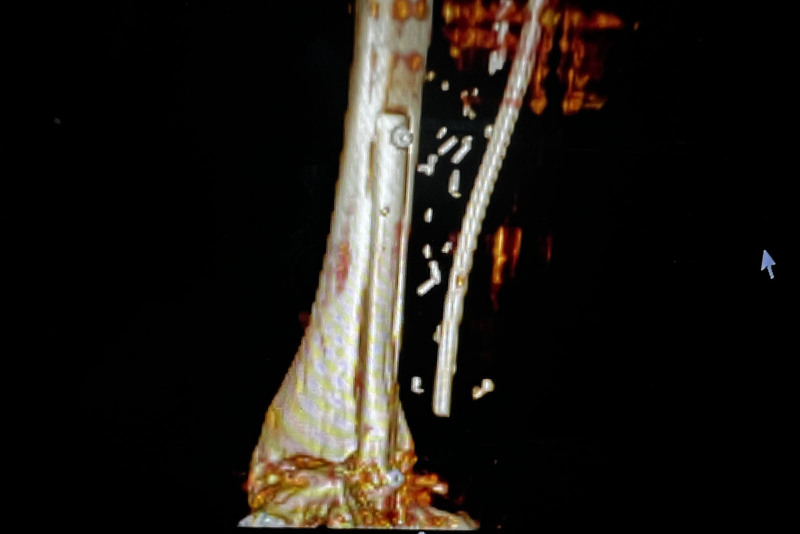
Results: A total of six patients were isolated, with three patients undergoing a free fibula approach and three patients undergoing the pedicled fibula approach. All patients were found to have tolerated the procedure well and had no intraoperative complications. In addition, all patients had clinically viable flaps and were satisfied with their surgical result.
Conclusions: Both free and pedicled free fibula flaps may be used effectively in the management of ankle arthrodesis in patients who have failed prior therapy. In our study, free fibula flaps were utilized in a medial approach, while the pedicled fibula flap was utilized in a lateral approach. With the right expertise and patient population, the free and pedicled fibula flaps can be highly successful in the repair of ankle defects.
Copyright © 2022 The Authors. Published by Wolters Kluwer Health, Inc. on behalf of The American Society of Plastic Surgeons.
Conflict of interest statement
Disclosure: The authors have no financial interest to declare in relation to the content of this article.
Figures


References
-
- Carlsson AS, Montgomery F, Besjakov J. Arthrodesis of the ankle secondary to replacement. Foot Ankle Int. 1998;19:240–245. - PubMed
-
- Albert E. Zur Resektion des Kniegelenkes. Wien; Med Press. 1879;20:705–708.
-
- Mendicino SS, Kreplick AL, Walters JL. Open ankle arthrodesis. Clin Podiatr Med Surg. 2017;34:489–502. - PubMed
-
- Bauer G, Kinzl L. Arthrodesen des oberen sprunggelenks [arthrodesis of the ankle joint]. Orthopade. 1996;25:158–165. - PubMed
LinkOut - more resources
Full Text Sources