Review
. 2022 Nov 10:10:1047256.
doi: 10.3389/fcell.2022.1047256. eCollection 2022.

Non-muscle myosin II and the plasticity of 3D cell migration

Affiliations
Review

Non-muscle myosin II and the plasticity of 3D cell migration

James M Cowan et al. Front Cell Dev Biol. .

Abstract

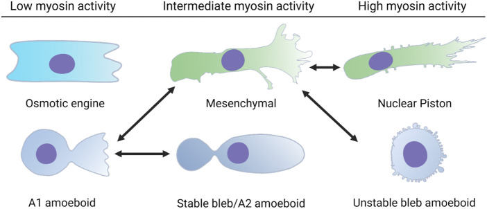
Confined cells migrating through 3D environments are also constrained by the laws of physics, meaning for every action there must be an equal and opposite reaction for cells to achieve motion. Fascinatingly, there are several distinct molecular mechanisms that cells can use to move, and this is reflected in the diverse ways non-muscle myosin II (NMII) can generate the mechanical forces necessary to sustain 3D cell migration. This review summarizes the unique modes of 3D migration, as well as how NMII activity is regulated and localized within each of these different modes. In addition, we highlight tropomyosins and septins as two protein families that likely have more secrets to reveal about how NMII activity is governed during 3D cell migration. Together, this information suggests that investigating the mechanisms controlling NMII activity will be helpful in understanding how a single cell transitions between distinct modes of 3D migration in response to the physical environment.

Keywords: cytoskeleton; matrix; mechanotranduction; motility; myosin.


Conflict of interest statement

The authors declare that the research was conducted in the absence of any commercial or financial relationships that could be construed as a potential conflict of interest.

Figures

FIGURE 1
FIGURE 1
Single cells can switch between distinct modes of 3D cell movement. Each of these modes of migration have different requirements for cell-matrix adhesion and actomyosin contractility. Cells using the osmotic engine or stable bleb modes of movement rely on friction against the surfaces that confine them to achieve forward movement. Cells using the unstable bleb amoeboid, 3D mesenchymal (lamellipodia-based), or nuclear piston (lobopodia-based) modes rely on cell-matrix adhesions to propel themselves forward. The double headed arrows indicate the known transitions that a single cell can use to switch between different modes of movement in response to the physical structure of the ECM and changes to intracellular signaling.
FIGURE 2
FIGURE 2
NMII activity powers multiple modes of 3D cell migration via distinct mechanisms. (A). Actomyosin contractility in the front of the cell is attached to vimentin intermediate filaments by the plectin crosslinker. The force generated by this machinery is transmitted to the nucleus via nesprin 3 to help pull the nucleus through 3D matrices. The forward movement of the nucleus increases cytoplasmic pressure to form lobopodial protrusions. (B). NMII activity at the trailing edge is regulated by mechanical stress on the nucleus, tension within the plasma membrane, and the small GTPase RhoA. This actomyosin contractility increases pressure at the rear of the cell and drives water into the nucleus which can lead to blebbing and rupture of the nuclear envelope. (C). NMII activity in leading protrusions can transmit traction forces to the surrounding ECM. This helps to align matrix fibers immediately in front of the cell to promote directional movement. (D). Actomyosin contractility in the cortex increases pressure in amoeboid cells forming small, unstable blebs. NMII-driven retrograde flow propels cells forming stable blebs forward through preexisting channels.
FIGURE 3
FIGURE 3
Tropomyosins, septins, and the BORG proteins can modulate the NMII activity within discrete populations of actin filaments. (A) Tpms can modulate the activity of NMII on F-actin. In this example, the association of Tpm 1.6 with actin stress fibers increases NMII activity. This mechanism aids in pressure-based 3D migration mechanisms such as the nuclear piston. (B) SEPT2 can promote actomyosin contractility through a direct interaction with NMII on F-actin. (C) BORG (binder of Rho GTPases) proteins are effectors of the small GTPase Cdc42 that bind and regulate septins to modulate intracellular force production by NMII.

References

    1. Adams G., Lopez M. P., Cartagena-Rivera A. X., Waterman C. M. (2021). Survey of cancer cell anatomy in nonadhesive confinement reveals a role for filamin-A and fascin-1 in leader bleb-based migration. Mol. Biol. Cell 32, 1772–1791. 10.1091/mbc.E21-04-0174 - DOI - PMC - PubMed
    1. Agarwal P., Zaidel-Bar R. (2019). Diverse roles of non-muscle myosin II contractility in 3D cell migration. Essays Biochem. 63, 497–508. 10.1042/EBC20190026 - DOI - PubMed
    1. Anderson T. W., Vaughan A. N., Cramer L. P. (2008). Retrograde flow and myosin II activity within the leading cell edge deliver F-actin to the lamella to seed the formation of graded polarity actomyosin II filament bundles in migrating fibroblasts. Mol. Biol. Cell 19, 5006–5018. 10.1091/mbc.e08-01-0034 - DOI - PMC - PubMed
    1. Appaduray M. A., Masedunskas A., Bryce N. S., Lucas C. A., Warren S. C., Timpson P., et al. (2016). Recruitment kinetics of tropomyosin Tpm3.1 to actin filament bundles in the cytoskeleton is independent of actin filament kinetics. PLoS One 11, e0168203. 10.1371/journal.pone.0168203 - DOI - PMC - PubMed
    1. Arbizzani F., Mavrakis M., Hoya M., Ribas J. C., Brasselet S., Paoletti A., et al. (2022). Septin filament compaction into rings requires the anillin Mid2 and contractile ring constriction. Cell Rep. 39, 110722. 10.1016/j.celrep.2022.110722 - DOI - PubMed

LinkOut - more resources