. 2022 Nov 9:9:1037941.
doi: 10.3389/fmolb.2022.1037941. eCollection 2022.

The mitochondrial Cu+ transporter PiC2 (SLC25A3) is a target of MTF1 and contributes to the development of skeletal muscle in vitro

Affiliations

The mitochondrial Cu+ transporter PiC2 (SLC25A3) is a target of MTF1 and contributes to the development of skeletal muscle in vitro

Cat McCann et al. Front Mol Biosci. .

Abstract

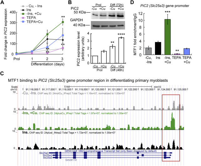
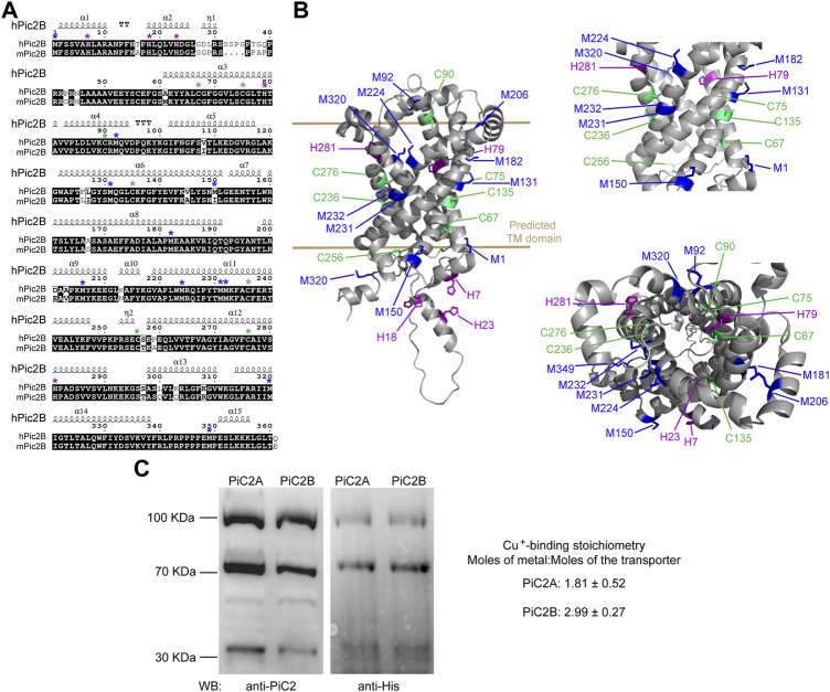
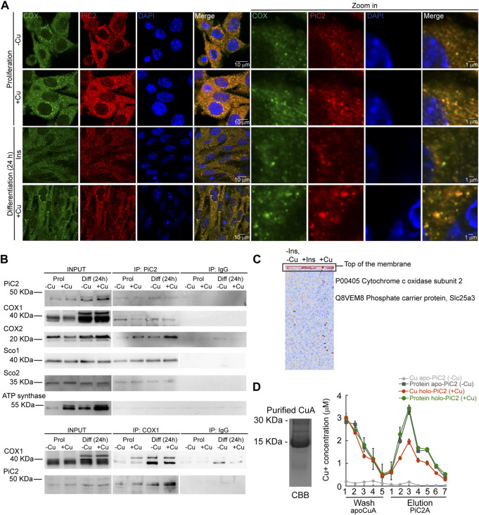
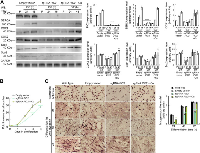
The loading of copper (Cu) into cytochrome c oxidase (COX) in mitochondria is essential for energy production in cells. Extensive studies have been performed to characterize mitochondrial cuproenzymes that contribute to the metallation of COX, such as Sco1, Sco2, and Cox17. However, limited information is available on the upstream mechanism of Cu transport and delivery to mitochondria, especially through Cu-impermeable membranes, in mammalian cells. The mitochondrial phosphate transporter SLC25A3, also known as PiC2, binds Cu+ and transports the ion through these membranes in eukaryotic cells, ultimately aiding in the metallation of COX. We used the well-established differentiation model of primary myoblasts derived from mouse satellite cells, wherein Cu availability is necessary for growth and maturation, and showed that PiC2 is a target of MTF1, and its expression is both induced during myogenesis and favored by Cu supplementation. PiC2 deletion using CRISPR/Cas9 showed that the transporter is required for proliferation and differentiation of primary myoblasts, as both processes are delayed upon PiC2 knock-out. The effects of PiC2 deletion were rescued by the addition of Cu to the growth medium, implying the deleterious effects of PiC2 knockout in myoblasts may be in part due to a failure to deliver sufficient Cu to the mitochondria, which can be compensated by other mitochondrial cuproproteins. Co-localization and co-immunoprecipitation of PiC2 and COX also suggest that PiC2 may participate upstream in the copper delivery chain into COX, as verified by in vitro Cu+-transfer experiments. These data indicate an important role for PiC2 in both the delivery of Cu to the mitochondria and COX, favoring the differentiation of primary myoblasts.

Keywords: MTF1; PiC2; SLC25A3; copper transport; cytochrome c oxidase; mitochondria.


Conflict of interest statement

The authors declare that the research was conducted in the absence of any commercial or financial relationships that could be construed as a potential conflict of interest.

Figures

FIGURE 1
FIGURE 1
Expression of mPiC2 increases over the course of differentiation in primary myoblasts derived from mouse satellite cells. (A) Representative confocal microscopy analysis of PiC2 expression and localization. Proliferating (48 h) and differentiating (24 h, 48 h, and 72 h) primary myoblasts were fixed, and stained with a fluorescent antibody against PiC2 (green) and DAPI (blue). (B) mPiC2 mRNA expression is induced during differentiation. qRT-PCR analysis of mPiC2 mRNA levels. Values normalized to Ef1α and non-differentiated (Prol) controls, n = 3. (C) mPiC2 protein expression is induced during differentiation. Representative western blot of PiC2 protein levels (top panel) and quantification (bottom panel). Membranes were blotted with PiC2, the differentiation marker myosin heavy chain (MHC), and GAPDH was used as loading control. n = 3 All values are reported as means ± SEM. Significance was determined by two-way ANOVA; *p < 0.05, **p < 0.01, ***p < 0.001 and ****p < 0.0001 compared to Prol cells.
FIGURE 2
FIGURE 2
mPic2 is a target gene of MTF1 in differentiating primary myoblasts, and Cu promotes this binding. (A) mRNA levels of mPiC2 in proliferating and differentiating primary myoblasts. mPic2 expression was measured by qPCR. Values normalized to Ef1α and non-differentiated (Prol) controls, n = 3. (B) Representative western blot (top) and quantification (bottom) of proliferating (48 h) and differentiating (72 h) primary myoblasts grown in the presence or absence of Cu. Values normalized to GAPDH and non-treated Prol controls, n = 3. (C) Genome browser tracks of ChIP-seq experiments assessing MTF1 binding to the mPic2 promoter. Promoter region of mPic2 shown in red box. (D) ChIP-qPCR validation of MTF1 binding the mPic2 promoter region. MTF1 enrichment at the mPic2 promoter was assessed by ChIP-qPCR, n = 3. All values are reported as means ± SEM. Significance was determined by two-way ANOVA; **p < 0.01 and ***p < 0.001 compared to cells differentiated with insulin. Abbreviations used are: cells differentiated in the absence of insulin and copper (-Ins, −Cu), with insulin (Ins), of insulin and 30 µM CuSO4 (-Ins, +Cu), with the 30 µM of the Cu-chelator TEPA (TEPA), and with 30 µM of TEPA supplemented with 30 µM CuSO4 (TEPA + Cu).
FIGURE 3
FIGURE 3
mPiC2 protein has several potential Cu binding sites. (A) Sequence homology comparison of the human hPiC2B and murine mPiC2B. Alignment of hPiC2B and mPiC2B amino acid sequences. Areas of homology between the sequences of the two transporters are indicated in black, potential Cu+-binding resides indicated with green asterisks for cysteines, blue asterisks for methionines and purple asterisks for histidines (*). (B) Representative model of mPic2 protein and potential Cu+-binding sites obtained from Alpha fold. Potential Cu+-binding residues indicated in green. (C) Representative western blots of recombinant PiC2A and PiC2B protein expression detected by both anti-PiC2 antibody and anti-His tag antibody. Monomeric, dimeric, and trimeric forms are detected by both antibodies. Cu+-binding stoichiometry of Pic2A and Pic2B. n = 3, results reported as means ± SEM.
FIGURE 4
FIGURE 4
mPiC2 interacts with cytochrome c oxidase and other mitochondrial cuproproteins in primary myoblasts derived from mouse satellite cells. (A) mPiC2 co-localizes with COX. Confocal imaging showing mPiC2 (red) and COX (green) co-localization (yellow). The nucleus is stained with DAPI (blue). Set of four panels on the right are zooms of areas in the set of four panels on the left. (B) Co-IP of mPiC2 with mitochondrial cuproproteins. Proliferating and differentiating primary myoblasts were treated with or without Cu. Co-IP was performed with and anti-PiC2 antibody and probed against PiC2 and COX1, COX2, Sco1, and Sco2; the ATP synthase was used as a negative control. Reciprocal co-IP using an anti-COX1 antibody was probed with an anti-COX1 and anti-PiC2 antibodies. (C) Synchrotron based X-Ray fluorescence analyses couple to protein sequencing by mass spectrometry of native PAGE gels from whole cell extracts of differentiating primary myoblasts showed the presence of mPiC2 and COX in a high molecular band that contained copper (indicated by black box). (D) Cu+ transfer from metallated PiC2A to apo-CuA. Cu+ concentration is shown in red, and the corresponding protein is shown in green. Control Cu+ transfer experiment is shown in pale (Cu+ signal) and dark gray (protein signal). In all cases, the contents of the wash and elution fractions are shown. The data presented in this figure corresponds to three independent biological replicates shown as the mean for Cu+ and protein concentration ±SEM.
FIGURE 5
FIGURE 5
Deletion of mPiC2 impairs the proliferation and differentiation of primary myoblasts derived from mouse satellite cells. (A) Deletion of mPiC2 reduces the expression of mitochondrial cuproproteins. Representative western blot of mPiC2 and mitochondrial cuproproteins in control cells (empty vector) or cells in which CRISPR/Cas9 was used to delete mPiC2 (sgRNA PiC2) or KO cells supplemented with CuSO4 (sgRNA PiC2 + Cu) during proliferation (P) and at 24 h and 48 h after inducing differentiation. GAPDH was used as a loading control. Plots represent the quantification of each protein analyzed from three independent biological replicates. Values are the means ± SEM. Significance was determined by two-way ANOVA; *p < 0.05, **p < 0.01 and ****p < 0.0001 vs. empty vector at the corresponding timepoint. (B) Deletion of mPiC2 delays proliferation. Cell counting assay of empty vector, sgRNA against PiC2, and sgRNA PiC2 + Cu cells at different points during proliferation. n = 3. All values are reported as means ± SEM. Significance was determined by two-way ANOVA; ***p < 0.001 and ****p < 0.0001 compared to empty vector cells. (C) Deletion of mPiC2 delays myogenesis. Staining of wild type, empty vector, sgRNA mPiC2, and sgRNA mPiC2 + Cu cells with either anti-Pax7 antibody (marker of proliferation) or anti-myogenin antibody (marker of differentiation). Associated plot represents the number of myogenin positive nuclei of cells undergoing differentiation at the indicated timepoints in the immunohistochemistry images n = 3. All values are reported as means ± SEM. Significance was determined by two-way ANOVA; ***p < 0.001 and ****p < 0.0001 compared to empty vector cells.

References

    1. Andrews G. K., Lee D. K., Ravindra R., Lichtlen P., Sirito M., Sawadogo M., et al. (2001). The transcription factors MTF-1 and USF1 cooperate to regulate mouse metallothionein-I expression in response to the essential metal zinc in visceral endoderm cells during early development. EMBO J. 20, 1114–1122. 10.1093/emboj/20.5.1114 - DOI - PMC - PubMed
    1. Argüello J. M., Raimunda D., Padilla-Benavides T. (2013). Mechanisms of copper homeostasis in bacteria. Front. Cell. Infect. Microbiol. 3, 73. 10.3389/fcimb.2013.00073 - DOI - PMC - PubMed
    1. Banci L., Bertini I., Ciofi-Baffoni S., Hadjiloi T., Martinelli M., Palumaa P. (2008). Mitochondrial copper(I) transfer from Cox17 to Sco1 is coupled to electron transfer. Proc. Natl. Acad. Sci. U. S. A. 105, 6803–6808. 10.1073/pnas.0800019105 - DOI - PMC - PubMed
    1. Banci L., Bertini I., Ciofi-Baffoni S., Janicka A., Martinelli M., Kozlowski H., et al. (2008). A structural-dynamical characterization of human Cox17. J. Biol. Chem. 283, 7912–7920. 10.1074/jbc.M708016200 - DOI - PubMed
    1. Banci L., Bertini I., Ciofi-Baffoni S., Kozyreva T., Zovo K., Palumaa P. (2010). Affinity gradients drive copper to cellular destinations. Nature 465, 645–648. 10.1038/nature09018 - DOI - PubMed