

Transplantation of neural stem progenitor cells from different sources for severe spinal cord injury repair in rat
- PMID: 36439085
- PMCID: PMC9692187
- DOI: 10.1016/j.bioactmat.2022.11.008
Transplantation of neural stem progenitor cells from different sources for severe spinal cord injury repair in rat
Abstract
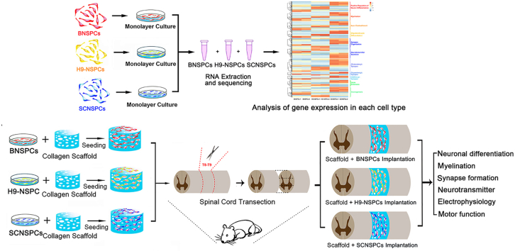
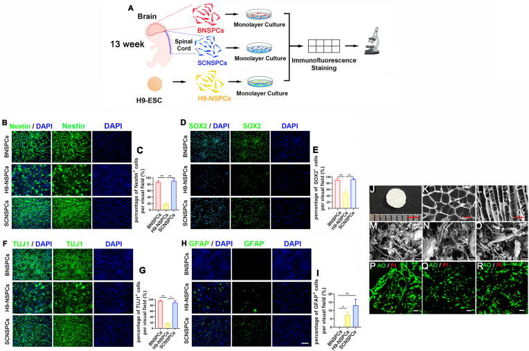
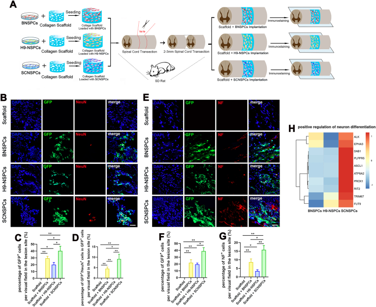
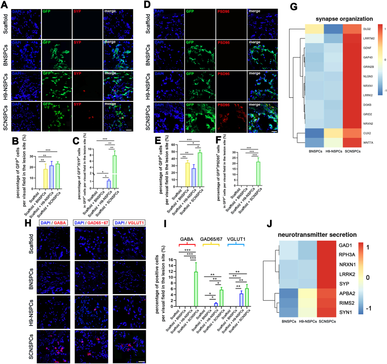
Neural stem progenitor cell (NSPC) transplantation has been regarded as a promising therapeutic method for spinal cord injury (SCI) repair. However, different NSPCs may have different therapeutic effects, and it is therefore important to identify the optimal NSPC type. In our study, we compared the transcriptomes of human fetal brain-derived NSPCs (BNSPCs), spinal cord-derived NSPCs (SCNSPCs) and H9 embryonic stem-cell derived NSPCs (H9-NSPCs) in vitro and subsequently we transplanted each NSPC type on a collagen scaffold into a T8-9 complete SCI rat model in vivo. In vitro data showed that SCNSPCs had more highly expressed genes involved in nerve-related functions than the other two cell types. In vivo, compared with BNSPCs and H9-NSPCs, SCNSPCs exhibited the best therapeutic effects; in fact, SCNSPCs facilitated electrophysiological and hindlimb functional recovery. This study demonstrates that SCNSPCs may be an appropriate candidate cell type for SCI repair, which is of great clinical significance.
Keywords: Brain-derived NSPCs; Collagen scaffolds; H9 embryonic stem cell-derived NSPCs; Spinal cord injury; Spinal cord-derived NSPCs.
© 2022 The Authors.
Conflict of interest statement
The authors declare that they have no known competing financial interests or personal relationships that could have appeared to influence the work reported in this paper.
Figures


Similar articles
-
Transcriptomic and Functional Landscape of Adult Human Spinal Cord NSPCs Compared to iPSC-Derived Neural Progenitor Cells.Cells. 2025 Jan 7;14(2):64. doi: 10.3390/cells14020064. Cells. 2025. PMID: 39851491 Free PMC article.
-
Comparison between fetal spinal-cord- and forebrain-derived neural stem/progenitor cells as a source of transplantation for spinal cord injury.Dev Neurosci. 2004 Mar-Aug;26(2-4):275-87. doi: 10.1159/000082144. Dev Neurosci. 2004. PMID: 15711067
-
Aligned collagen scaffold combination with human spinal cord-derived neural stem cells to improve spinal cord injury repair.Biomater Sci. 2020 Sep 21;8(18):5145-5156. doi: 10.1039/d0bm00431f. Epub 2020 Aug 24. Biomater Sci. 2020. PMID: 32832944
-
hiPSC-Neural Stem/Progenitor Cell Transplantation Therapy for Spinal Cord Injury.Curr Stem Cell Res Ther. 2023;18(4):487-498. doi: 10.2174/1574888X17666220509222520. Curr Stem Cell Res Ther. 2023. PMID: 35538805 Review.
-
Review of transplantation of neural stem/progenitor cells for spinal cord injury.Int J Dev Neurosci. 2013 Nov;31(7):701-13. doi: 10.1016/j.ijdevneu.2013.07.004. Epub 2013 Aug 6. Int J Dev Neurosci. 2013. PMID: 23928260 Review.
Cited by
-
The activation of dormant ependymal cells following spinal cord injury.Stem Cell Res Ther. 2023 Jul 5;14(1):175. doi: 10.1186/s13287-023-03395-4. Stem Cell Res Ther. 2023. PMID: 37408068 Free PMC article. Review.
-
Conjugated therapy with coaxially printed neural stem cell-laden microfibers and umbilical cord mesenchymal stem cell derived exosomes on complete transactional spinal cord defects.Mater Today Bio. 2025 Mar 4;32:101639. doi: 10.1016/j.mtbio.2025.101639. eCollection 2025 Jun. Mater Today Bio. 2025. PMID: 40160243 Free PMC article.
-
Bibliometric analysis of stem cells for spinal cord injury: current status and emerging frontiers.Front Pharmacol. 2023 Jul 18;14:1235324. doi: 10.3389/fphar.2023.1235324. eCollection 2023. Front Pharmacol. 2023. PMID: 37533634 Free PMC article.
-
Targeting Active Microglia Alleviates Distal Edge of Proton Radiation-induced Neural Damage.Adv Radiat Oncol. 2025 Mar 18;10(5):101764. doi: 10.1016/j.adro.2025.101764. eCollection 2025 May. Adv Radiat Oncol. 2025. PMID: 40291513 Free PMC article.
-
Transcriptomic and Functional Landscape of Adult Human Spinal Cord NSPCs Compared to iPSC-Derived Neural Progenitor Cells.Cells. 2025 Jan 7;14(2):64. doi: 10.3390/cells14020064. Cells. 2025. PMID: 39851491 Free PMC article.
References
-
- McDonald J.W., Sadowsky C. Spinal-cord injury. Lancet. 2002;359(9304):417–425. - PubMed
-
- Assinck P., Duncan G.J., Hilton B.J., Plemel J.R., Tetzlaff W. Cell transplantation therapy for spinal cord injury. Nat. Neurosci. 2017;20(5):637–647. - PubMed
-
- Ostenfeld T., Joly E., Tai Y.T., Peters A., Caldwell M., Jauniaux E., Svendsen C.N. Regional specification of rodent and human neurospheres. Dev. Brain Res. 2002;134(1–2):43–55. - PubMed
-
- Horiguchi S., Takahashi J., Kishi Y., Morizane A., Okamoto Y., Koyanagi M., Tsuji M., Tashiro K., Honjo T., Fujii S., Hashimoto N. Neural precursor cells derived from human embryonic brain retain regional specificity. J. Neurosci. Res. 2004;75(6):817–824. - PubMed
LinkOut - more resources
Full Text Sources
