. 2022 Nov 10:13:994626.
doi: 10.3389/fimmu.2022.994626. eCollection 2022.

Dorsal switch protein 1 as a damage signal in insect gut immunity to activate dual oxidase via an eicosanoid, PGE2

Affiliations

Dorsal switch protein 1 as a damage signal in insect gut immunity to activate dual oxidase via an eicosanoid, PGE2

Miltan Chandra Roy et al. Front Immunol. .

Abstract

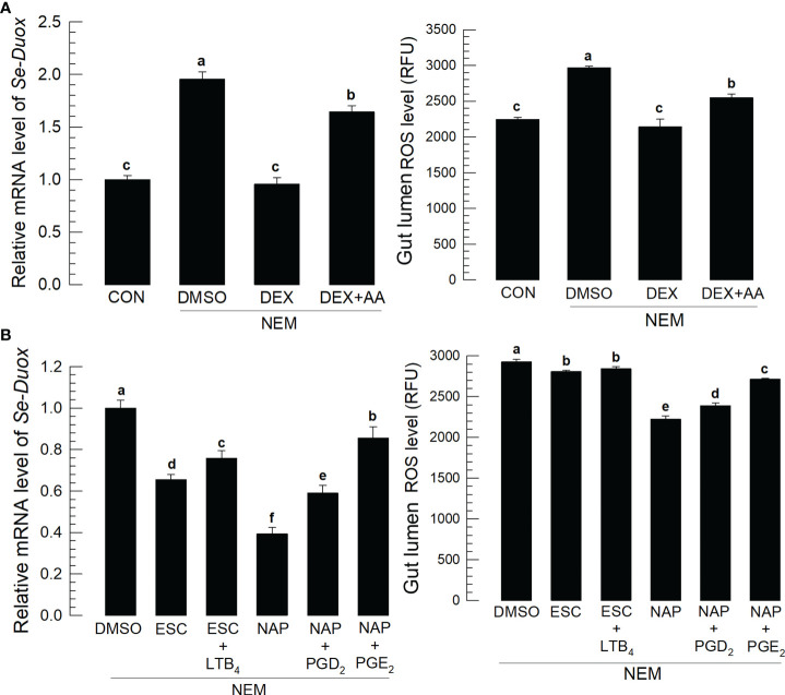
Various microbiota including beneficial symbionts reside in the insect gut. Infections of pathogens cause dysregulation of the microflora and threaten insect survival. Reactive oxygen species (ROS) have been used in the gut immune responses, in which its production is tightly regulated by controlling dual oxidase (Duox) activity via Ca2+ signal to protect beneficial microflora and gut epithelium due to its high cytotoxicity. However, it was not clear how the insects discriminate the pathogens from the various microbes in the gut lumen to trigger ROS production. An entomopathogenic nematode (Steinernema feltiae) infection elevated ROS level in the gut lumen of a lepidopteran insect, Spodoptera exigua. Dorsal switch protein 1 (DSP1) localized in the nucleus in the midgut epithelium was released into plasma upon the nematode infection and activated phospholipase A2 (PLA2). The activated PLA2 led to an increase of PGE2 level in the midgut epithelium, in which rising Ca2+ signal up-regulated ROS production. Inhibiting DSP1 release by its specific RNA interference (RNAi) or specific inhibitor, 3-ethoxy-4-methoxyphenol, treatment failed to increase the intracellular Ca2+ level and subsequently prevented ROS production upon the nematode infection. A specific PLA2 inhibitor treatment also prevented the up-regulation of Ca2+ and subsequent ROS production upon the nematode infection. However, the addition of PGE2 to the inhibitor treatment rescued the gut immunity. DSP1 release was not observed at infection with non-pathogenic pathogens but detected in plasma with pathogenic infections that would lead to damage to the gut epithelium. These results indicate that DSP1 acts as a damage-associated molecular pattern in gut immunity through DSP1/PLA2/Ca2+/Duox.

Keywords: DSP1; PGE2; eicosanoid; gut; immunity; insect.


Conflict of interest statement

The authors declare that the research was conducted in the absence of any commercial or financial relationships that could be construed as a potential conflict of interest.

Figures

Figure 1
Figure 1
Entomopathogenic nematode (‘NEM’) infection to S. exigua gut leads to ROS burst. Each L5 larva of S. exigua was fed with 80 IJs of S. feltiae. (A) Temporal IJ infection from gut lumen to hemocoel after oral feeding treatment. (B) Induction of Se-Duox expression and ROS level. (C) Modulation of ROS levels against the nematode infection and subsequent virulence to S. exigua. L5 larvae were infected with IJs at 24 h after vitamin C (‘Vit-C’, 100 mg/larva) and paraquat (25 µg/larva) treatment. Larval mortality was recorded 72 h after the nematode treatment. Each treatment was replicated three times. Different letters and asterisks above standard error bars indicate significant differences among means at Type I error = 0.05 (LSD test). NS, no significance.
Figure 2
Figure 2
Release of Se-DSP1 from the midgut epithelium to defend the entomopathogenic nematode (‘NEM’) infection. Each L5 larva of S. exigua was fed with 80 IJs of S. feltiae. (A) Nuclear localization of Se-DSP1 in the midgut epithelium of S. exigua. An immunofluorescence image was obtained by staining Se-DSP1 using a specific antibody labeled with FITC and nuclei with DAPI. ‘DIC’ represents differential interference contrast. The scale bar represents 10 µm. (B) A western blotting showing release of Se-DSP1 upon the nematode infection into plasma, which was obtained 8 h after the nematode treatment. 3-Ethoxy-4-methoxyphenol (‘EMP’, 1,000 ppm) was treated along with the nematodes. α-Tubulin was stained as a control to confirm the equal amount of protein loading. (C) Effect of Se-DSP1 release on PLA2 activity in plasma, Se-Duox expression in the epithelium, and ROS level in the gut lumen. (D) Enhancement of the nematode (80 IJs/larva) virulence with the addition of EMP against L5 larvae of S. exigua. Mortality was assessed at 48 h after the nematode treatment. (E) Effect of PLA2 activity induced by Se-DSP1 release on Se-Duox expression and ROS production. RNAi was performed by injecting dsRNA (1 µg) specific to Se-DSP1. The nematode treatment was followed at 24 h after the dsRNA injection. At 8 h after the nematode treatment, Se-Duox expression and ROS production were assessed. Each treatment was replicated three times. Different letters above the standard error bars indicate significant differences among means at Type I error = 0.05 (LSD test).
Figure 3
Figure 3
Effects of eicosanoids on expression levels of Se-Duox and ROS production in response to gut infection by the entomopathogenic nematode (‘NEM’). Each L5 larva of S. exigua was fed with 80 IJs of S. feltiae. (A) Inhibitory effect of a PLA2 inhibitor, dexamethasone (‘DEX’), on Se-Duox expression and ROS level in the gut lumen of L5 larvae. NEM treatment was performed 8 h post-injection (PI) of DEX (10 µg/larva) or arachidonic acid (AA, 10 µg/larva). At 8 h after NEM treatment, Se-Duox expression and ROS level were measured. (B) Effect of naproxene (‘NAP’, a COX inhibitor) and esculetin (‘ESC’, a LOX inhibitor) on the expression of Se-Duox and ROS level. To rescue the inhibitor treatments, LTB4, PGD2, or PGE2 was injected at 1 μg/larva along with ESC or NAP treatment. Each treatment was replicated three times. Different letters above the standard error bars indicate significant differences among means at Type I error = 0.05 (LSD test).
Figure 4
Figure 4
Ca2+ signal after Se-DSP1 release via PGE2 in response to gut infection by the entomopathogenic nematode (‘NEM’). Each L5 larva of S. exigua was fed with 80 IJs of S. feltiae. (A) Ca2+ signals in the entire gut are separated into anterior (‘A’), central (‘C’), and posterior (‘P’). Nuclei were stained with DAPI. Fura-8AM was used to observe Ca2+. ‘DIC’ represents differential interference contrast. The scale bar indicates 100 µm. (B) Induction of Ca2+ signal and subsequent up-regulation of ROS level in midgut in response to NEM treatment for 8 h. (C) Effect of RNAi specific to Se-DSP1 expression on Ca2+ signal in response to NEM treatment. At 24 h after dsRNA (1 µg/larva) injection, NEM was treated. After 8 h NEM treatment, Ca2+ level and ROS amount were measured. (D) Induction of PGE2 in the midgut epithelium. (E) Inhibitory effect of a specific PLA2 inhibitor (dexamethasone (DEX) on Ca2+ signal and ROS level in midgut. NEM treatment was performed at 8 h after injection of DEX (10 µg/larva) or PGE2 (1 µg/larva). Nuclei were stained with DAPI. PGE2 was detected with a specific antibody raised against rabbit. Fura-8AM was used to observe Ca2+. Scale bars in B-E represent 10 µm. Different letters above standard error bars indicate significant difference among means at Type I error = 0.05 (LSD test).
Figure 5
Figure 5
Effect of PGE2 receptor (‘PGE2R’) and its downstream signal on the induction of Ca2+ signal in response to gut infection by the entomopathogenic nematode (‘NEM’) infection. Each L5 larva of S. exigua was fed with 80 IJs of S. feltiae. (A) Effect of RNAi against Se-PGE2R expression on calcium signal and ROS production. NEM treatment was performed at 24 h after dsRNA (1 µg/larva) injection. (B) Inhibition of Ca2+ signal by four different calcium signal inhibitors: dantrolene sodium (‘DAN’, a specific inhibitor to ryanodine receptor), 2-APB (a specific IP3 receptor inhibitor), U-73122 (a specific PLC inhibitor) along with NEM treatment. All inhibitors were injected in a dose of 1 µg per larva. The guts of treated larvae were dissected at 8 h after the NEM or inhibitor treatment. Nuclei were stained with DAPI. Fura-8AM was used to observe Ca2+. Each treatment was replicated three times. The scale bar represents 10 µm. Different letters above standard error bars indicate significant difference among means at Type I error = 0.05 (LSD test).
Figure 6
Figure 6
Release of Se-DSP1 into plasma upon infection of different microbes. Western blotting shows the release of Se-DSP1 into plasma upon gut infection by (A) entomopathogenic fungus, M. rileyi (‘Mr’), entomopathogenic bacterium, B. thuringiensis subsp. aizawai (‘BtA’), a baculovirus (‘SeMNPV’), and (B) non-pathogenic bacteria, E. coli (‘Ec’) and P. agglomerans (‘Pa’). Hemolymph was collected and plasma was separated at 0-48 h PI. Se-DSP1 antibody was used to detect DSP1 in plasma where α-tubulin was used as a reference to confirm the equal amount of protein loading.
Figure 7
Figure 7
A model of PGE2 mediating DSP1/Ca2+/Duox signaling pathway in the midgut epithelium of S. exigua in response to an entomopathogenic nematode (‘NEM’) infection. Upon infection of gut epithelium by EPN, a damage-associated pattern molecule (‘DSP1’) is released to hemocoel. Released DSP1 binds to spätzle 1 (‘Spz1’) to activate the Toll9 receptor to produce PGE2 in the gut epithelium or fat body (‘FB’) by activating phospholipase A2 (‘PLA2’) [20]. The autocrine/paracrine PGE2 binds to its specific receptor (‘PGE2R’) to up-regulate cAMP, which triggers Se-Duox expression via PKA/CREB [15]. cAMP also activates phospholipase C (‘PLC’) to increase the inositol triphosphate (‘IP3’) level [19]. IP3 binds to its receptor (‘IP3R’) on the endoplasmic reticulum to release Ca2+ [19]. The released Ca2+ triggers calcium-induced calcium release from the ryanodine receptor (‘RyR’), which leads to a Ca2+ burst [19]. The up-regulated Ca2+ then activates dual oxidase (‘Duox’) to produce reactive oxygen species (‘ROS’) and finally defend NEM.

Similar articles

Cited by

References

    1. Zhang M, Ma P, Zhang T, Gao Z, Zhao P, Liu X, et al. . Roles of LmCDA1 and LmCDA2 in cuticle formation in the foregut and hindgut of locusta migratoria. Insect Sci (2021) 28:1314–25. doi: 10.1111/1744-7917.12874 - DOI - PubMed
    1. Wu K, Yang B, Huang W, Dobens L, Song H, Ling E. Gut immunity in lepidopteran insects. Dev Comp Immunol (2016) 64:65–74. doi: 10.1016/j.dci.2016.02.010 - DOI - PubMed
    1. Hou D, Kuang W, Luo S, Zhang F, Zhou F, Chen T, et al. . Baculovirus ODV-E66 degrades larval peritrophic membrane to facilitate baculovirus oral infection. Virology (2019) 537:157–64. doi: 10.1016/j.virol.2019.08.027 - DOI - PubMed
    1. Leonard SP, Powell JE, Perutka J, Geng P, Heckmann LC, Horak RD, et al. . Engineered symbionts activate honey bee immunity and limit pathogens. Science (2020) 367:573–6. doi: 10.1126/science.aax9039 - DOI - PMC - PubMed
    1. Ryu JH, Kim SH, Lee HY, Bai JY, Nam YD, Bae JW, et al. . Innate immune homeostasis by the homeobox gene caudal and commensal-gut mutualism in drosophila. Science (2008) 319:777–82. doi: 10.1126/science.1149357 - DOI - PubMed

Publication types