. 2022 Nov 9:13:1023255.
doi: 10.3389/fimmu.2022.1023255. eCollection 2022.

Non-replicative antibiotic resistance-free DNA vaccine encoding S and N proteins induces full protection in mice against SARS-CoV-2

Affiliations

Non-replicative antibiotic resistance-free DNA vaccine encoding S and N proteins induces full protection in mice against SARS-CoV-2

Pedro J Alcolea et al. Front Immunol. .

Abstract

SARS-CoV-2 vaccines currently in use have contributed to controlling the COVID-19 pandemic. Notwithstanding, the high mutation rate, fundamentally in the spike glycoprotein (S), is causing the emergence of new variants. Solely utilizing this antigen is a drawback that may reduce the efficacy of these vaccines. Herein we present a DNA vaccine candidate that contains the genes encoding the S and the nucleocapsid (N) proteins implemented into the non-replicative mammalian expression plasmid vector, pPAL. This plasmid lacks antibiotic resistance genes and contains an alternative selectable marker for production. The S gene sequence was modified to avoid furin cleavage (Sfs). Potent humoral and cellular immune responses were observed in C57BL/6J mice vaccinated with pPAL-Sfs + pPAL-N following a prime/boost regimen by the intramuscular route applying in vivo electroporation. The immunogen fully protected K18-hACE2 mice against a lethal dose (105 PFU) of SARS-CoV-2. Viral replication was completely controlled in the lungs, brain, and heart of vaccinated mice. Therefore, pPAL-Sfs + pPAL-N is a promising DNA vaccine candidate for protection from COVID-19.

Keywords: DNA vaccine; N protein; S protein; SARS-CoV-2; furin; mouse model; pPAL.


Conflict of interest statement

The authors declare that the research was conducted in the absence of any commercial or financial relationships that could be construed as a potential conflict of interest.

Figures

Figure 1
Figure 1
The pPAL-Sfs + pPAL-N vaccine candidate. (A) pPAL-Sfs map. Sfs is a codon-optimized modified version of the SARS-CoV-2 Wuhan reference sequence that contains a modification to avoid furin cleavage of the protein product. Large-scale laboratory preparation of the pPAL-Sfs plasmid containing supercoiled (CCC), open-circular (OC), CCC-dimer, and CCC-trimer conformers. Western blot of whole protein extracts from HEK293 cells transfected with pPAL, pPAL-S, and pPAL-Sfs. The primary antibody was 1:800-diluted goat anti-S polyclonal antibody (Abcam #ab272504, Cambridge, United Kingdom), which only recognizes the S2 subunit. The secondary antibody was 1:2,000-diluted HRP-conjugated goat polyclonal anti-rabbit Ig. Furin cleaves the S protein obtained from pPAL-S but not Sfs from pPAL-Sfs. (B) Large-scale laboratory preparation of the pPAL-N plasmid containing CCC, OC, CCC-dimer, and CCC-trimer conformers. Western blot of whole protein extracts from HEK293 cells transfected with pPAL and pPAL-N. The primary antibody was 1:500-diluted rabbit anti-N polyclonal antibody. The secondary antibody was 1:2,000-diluted HRP-conjugated goat polyclonal anti-rabbit Ig. (C, D) immunofluorescence of cultured HEK293 cells transfected with pPAL, pPAL-Sfs, and pPAL-N plasmids. The anti-S and anti-N antibody dilutions were 1:50. The pPAL control was incubated with both antibodies. The secondary antibody was Alexa Fluor® 488-conjugated goat anti-rabbit IgG diluted to 1:200. Sfs and N expression is observed.
Figure 2
Figure 2
The pPAL-Sfs + pPAL-N vaccine candidates activate SARS-CoV-2 S- and RBD-specific humoral immunity in C57BL/6J mice. The serum samples were collected 15 days after the booster dose. (A) ELISA determinations of individual and average titers of total circulating anti-S and anti-RBD IgG determined by ELISA using HRP-conjugated protein (A) ELISA determinations of individual and average titers of circulating anti-RBD IgG, IgG1, and IgG2c using HRP-conjugated goat anti-mouse IgG1, anti-IgG2, and anti-IgG2c. The average anti-S and anti-RBD IgG titers are ≥2,000. The IgG2c/IgG1 ratio is ~750 in vaccinated animals with respect to the pPAL control group. (B) The neutralizing antibody titer average is >100. Statistical inference was performed using the Student’s t-test applying p-value adjustment by the Holm-Sidak method (α = 0.05; *p<0.05; **p<0.01; ***p<0.001; ns, non-significant).
Figure 3
Figure 3
S- and N-specific activation of IFN-γ-producing splenocytes induced by the pPAL-Sfs + pPAL-N vaccine candidate in C57BL/6J mice is dose-dependent. Splenocytes were obtained 7 days post-boost after prime/boost inoculation of pPAL and pPAL-Sfs + pPAL-N. Splenocytes (2x10⁵ cells/well) were stimulated at 37 °C in IFN-γ ELISpot plates for 16 h with 11-mer 8 amino acid-overlap peptide pools (25 µg/well) representing the whole S and N amino acid sequences in equimolar amounts. Negative stimulation controls were subtracted from the SFC per million cell values. Statistical inference was performed using the Student’s t-test applying p-value adjustment by the Holm-Sidak method (α = 0.05; **p<0.01; ***p<0.001). (A) Robust S- and N-specific activation of IFN-γ-producing splenocytes is observed with a prime/boost immunization regime with pPAL-Sfs + pPAL-N. (B) S-specific activation of IFN-γ-producing splenocytes with pPAL-Sfs + pPAL-N is dose dependent.
Figure 4
Figure 4
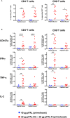
Specific CD3+CD4+ and CD3+CD8+ T-cell response induced by pPAL-Sfs + pPAL-N in C57BL/6J mice. Splenocytes were obtained from mice after prime/boost inoculation with pPAL and pPAL-Sfs + pPAL-N. Splenocytes were stimulated for 4 h with 11-mer 8 amino acid-overlap peptide pools representing the whole S and N amino acid sequences in equimolar amounts. (A) Percentage of CD3+CD4+ and CD3+CD8+ T cells producing CD107a, IFN-γ, TNF-α, and IL-2 in response to stimulation with peptide S or N pools. (B) Percentage of CD4+ and CD8+ T cells responding to S and N peptide pools above background, calculated as the sum of T cells positive for CD107a and/or IFN-γ and/or TNF-α and/or IL-2. Statistical inference using Two-way ANOVA with Fisher’s LD post-hoc test (*p<0.05; **p<0.01; ***p<0.001).
Figure 5
Figure 5
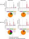
Polyfunctionality analysis of specific CD3+CD4+ and CD3+CD8+ T cell populations stimulated by pPAL-Sfs + pPAL-N immunization in C57BL/6J mice. Analyses of concomitant expression of CD107a, IFN-γ, TNF-α, and IL-2 in CD3+CD4+ T cells and CD3+CD8+ T cells in response to N peptide pool or S peptide pool stimulation. Pie charts split the percentage of T cells expressing either 1, 2, 3, or 4 markers upon stimulation. Statistical inference using Two-way ANOVA with Fisher’s LD post-hoc test (*p<0.05; **p<0.01; ***p<0.001).
Figure 6
Figure 6
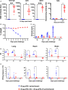
The pPAL-Sfs + pPAL-N vaccine confers full protection against challenge with 105 PFU of the MAD6 SARS-CoV-2 isolate in the K18-hACE2 murine model. (A) Titration of circulating anti-RBD IgG, IgG1, IgG2c, and neutralizing antibodies 15 days after prime/boost immunization with pPAL-Sfs + pPAL-N. (B) Body weight evolution after 105 PFU SARS-CoV-2 challenge. Vaccinated animals maintained their weight throughout the experiment. Weight decreased in the control group until the endpoint criteria had to be applied (20% of weight loss). (C) Clinical sign follow-up after challenge. (D) Reduction of the viral load in target organs. ΔCt accounting for SARS-CoV-2 mRNA levels. β-actin was the reference gene. (E) Reduction of viral replication (PFU/g tissue) in VERO cells. Statistical inference was performed using the Student’s t-test applying p-value adjustment by the Holm-Sidak method (α = 0.05; *p<0.05; **p<0.01; ***p<0.001; ****p<0.0001).
Figure 7
Figure 7
Full protection of K18-hACE2 mice after three months of vaccination with pPAL-Sfs+ pPAL-N against challenge with 105 PFU of the MAD6 SARS-CoV-2 isolate in the K18-hACE2 murine model. (A) Titration of circulating anti-RBD IgG, IgG1, and IgG2c three months after the pPAL-Sfs + pPAL-N booster dose administration. (B) Body weight evolution after 105 PFU SARS-CoV-2 challenge. Vaccinated animals maintained their weight throughout the experiment, whereas weight decreased in the control group. (C) Clinical sign follow-up after challenge with 105 PFU SARS-CoV-2. Statistical inference was performed using the Student’s t-test applying p-value adjustment by the Holm-Sidak method (α = 0.05; *p<0.05; **p<0.01).
Figure 8
Figure 8
The pPAL-Sfs + pPAL-N vaccine confers full protection against challenge with 105 PFU of the SARS-CoV-2 Delta variant in the K18-hACE2 murine model. (A) Titration of circulating anti-RBD IgG, IgG2c, and neutralizing antibodies 15 days after prime/boost immunization with pPAL-Sfs + pPAL-N. (B) Body weight evolution after 105 PFU SARS-CoV-2 challenge. Vaccinated animals maintained their weight throughout the experiment, whereas weight decreased in the control group. (C) Clinical sign follow-up after challenge with 105 PFU SARS-CoV-2. Statistical inference was performed using the Student’s t-test applying p-value adjustment by the Holm-Sidak method (α = 0.05; *p<0.05; ****p<0.0001).

References

    1. Richner JM, Himansu S, Dowd KA, Butler SL, Salazar V, Fox JM, et al. . Modified mRNA vaccines protect against zika virus infection. Cell (2017) 169(1):176. doi: 10.1016/j.cell.2017.02.017 - DOI - PubMed
    1. Pardi N, Hogan MJ, Pelc RS, Muramatsu H, Andersen H, DeMaso CR, et al. . Zika virus protection by a single low-dose nucleoside-modified mRNA vaccination. Nature (2017) 543(7644):248–51. doi: 10.1038/nature21428 - DOI - PMC - PubMed
    1. Reynolds CJ, Gibbons JM, Pade C, Lin KM, Sandoval DM, Pieper F, et al. . Heterologous infection and vaccination shapes immunity against SARS-CoV-2 variants. Science (2022) 375(6577):183–92. doi: 10.1126/science.abm0811 - DOI - PMC - PubMed
    1. Muik A, Lui BG, Wallisch AK, Bacher M, Muhl J, Reinholz J, et al. . Neutralization of SARS-CoV-2 omicron by BNT162b2 mRNA vaccine-elicited human sera. Science (2022) 375(6581):678–80. doi: 10.1126/science.abn7591 - DOI - PMC - PubMed
    1. Mannar D, Saville JW, Zhu X, Srivastava SS, Berezuk AM, Tuttle KS, et al. . SARS-CoV-2 omicron variant: Antibody evasion and cryo-EM structure of spike protein-ACE2 complex. Science (2022) 375(6582):760–4. doi: 10.1126/science.abn7760 - DOI - PMC - PubMed

Publication types