

The implication of a diversity of non-neuronal cells in disorders affecting brain networks
- PMID: 36439206
- PMCID: PMC9693782
- DOI: 10.3389/fncel.2022.1015556
The implication of a diversity of non-neuronal cells in disorders affecting brain networks
Abstract
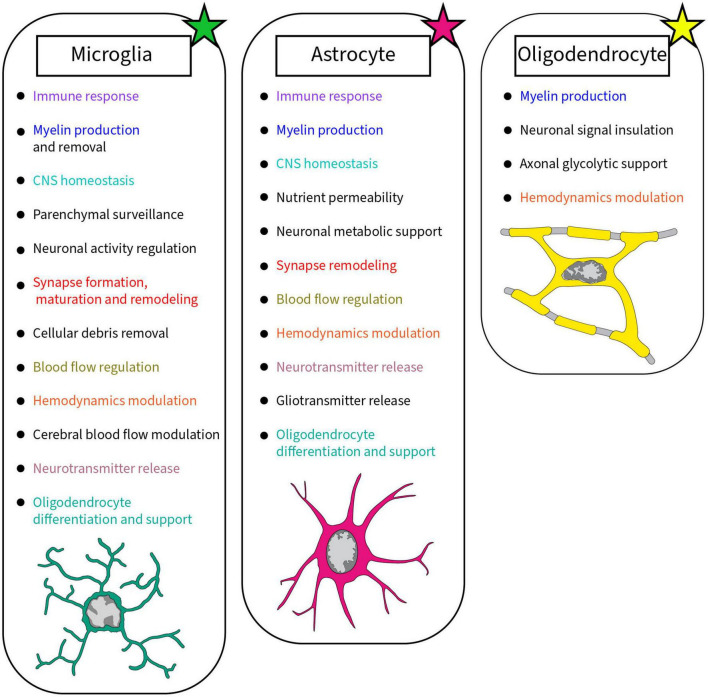
In the central nervous system (CNS) neurons are classically considered the functional unit of the brain. Analysis of the physical connections and co-activation of neurons, referred to as structural and functional connectivity, respectively, is a metric used to understand their interplay at a higher level. A myriad of glial cell types throughout the brain composed of microglia, astrocytes and oligodendrocytes are key players in the maintenance and regulation of neuronal network dynamics. Microglia are the central immune cells of the CNS, able to affect neuronal populations in number and connectivity, allowing for maturation and plasticity of the CNS. Microglia and astrocytes are part of the neurovascular unit, and together they are essential to protect and supply nutrients to the CNS. Oligodendrocytes are known for their canonical role in axonal myelination, but also contribute, with microglia and astrocytes, to CNS energy metabolism. Glial cells can achieve this variety of roles because of their heterogeneous populations comprised of different states. The neuroglial relationship can be compromised in various manners in case of pathologies affecting development and plasticity of the CNS, but also consciousness and mood. This review covers structural and functional connectivity alterations in schizophrenia, major depressive disorder, and disorder of consciousness, as well as their correlation with vascular connectivity. These networks are further explored at the cellular scale by integrating the role of glial cell diversity across the CNS to explain how these networks are affected in pathology.
Keywords: astrocytes; microglia; neurons; oligodendrocytes; structural and functional connectivity; synapses; vasculature.
Copyright © 2022 Carrier, Dolhan, Bobotis, Desjardins and Tremblay.
Conflict of interest statement
The authors declare that the research was conducted in the absence of any commercial or financial relationships that could be construed as a potential conflict of interest.
Figures
Similar articles
-
Neuroglia - development and role in physiological and pathophysiological processes.Folia Morphol (Warsz). 2021;80(4):766-775. doi: 10.5603/FM.a2021.0109. Epub 2021 Oct 26. Folia Morphol (Warsz). 2021. PMID: 34699052 Review.
-
Traumatic Brain Injury: Mechanisms of Glial Response.Front Physiol. 2021 Oct 22;12:740939. doi: 10.3389/fphys.2021.740939. eCollection 2021. Front Physiol. 2021. PMID: 34744783 Free PMC article. Review.
-
The Structure and Function of Glial Networks: Beyond the Neuronal Connections.Neurosci Bull. 2023 Mar;39(3):531-540. doi: 10.1007/s12264-022-00992-w. Epub 2022 Dec 8. Neurosci Bull. 2023. PMID: 36481974 Free PMC article. Review.
-
Astrocytes and microglia in the coordination of CNS development and homeostasis.J Neurochem. 2024 Oct;168(10):3599-3614. doi: 10.1111/jnc.16006. Epub 2023 Nov 20. J Neurochem. 2024. PMID: 37985374 Review.
-
Neuronal injury in chronic CNS inflammation.Best Pract Res Clin Anaesthesiol. 2010 Dec;24(4):551-62. doi: 10.1016/j.bpa.2010.11.001. Epub 2010 Nov 29. Best Pract Res Clin Anaesthesiol. 2010. PMID: 21619866 Review.
Cited by
-
Established and emerging techniques for the study of microglia: visualization, depletion, and fate mapping.Front Cell Neurosci. 2024 Feb 15;18:1317125. doi: 10.3389/fncel.2024.1317125. eCollection 2024. Front Cell Neurosci. 2024. PMID: 38425429 Free PMC article. Review.
-
Microglial responses to inflammatory challenge in adult rats altered by developmental exposure to polychlorinated biphenyls in a sex-specific manner.Neurotoxicology. 2024 Sep;104:95-115. doi: 10.1016/j.neuro.2024.07.009. Epub 2024 Jul 20. Neurotoxicology. 2024. PMID: 39038526
-
Associations among plasma markers for N-methyl-d-aspartate receptor hypofunction, redox dysregulation, and insufficient myelination in patients with schizophrenia.Heliyon. 2024 Apr 25;10(9):e30193. doi: 10.1016/j.heliyon.2024.e30193. eCollection 2024 May 15. Heliyon. 2024. PMID: 38694089 Free PMC article.
-
Synapse Regulation.Adv Neurobiol. 2024;37:179-208. doi: 10.1007/978-3-031-55529-9_11. Adv Neurobiol. 2024. PMID: 39207693 Review.
References
Publication types
LinkOut - more resources
Full Text Sources