. 2022 Nov 10:9:1042184.
doi: 10.3389/fsurg.2022.1042184. eCollection 2022.

Endoscopic lumbar foraminotomy for foraminal stenosis in stable spondylolisthesis

Affiliations

Endoscopic lumbar foraminotomy for foraminal stenosis in stable spondylolisthesis

Yong Ahn et al. Front Surg. .

Abstract

Background: Open decompression with fusion is the gold-standard surgical technique for spondylolisthesis. However, it may be too extensive for patients with foraminal stenosis with stable spondylolisthesis. The endoscopic lumbar foraminotomy (ELF) technique was developed as a minimally invasive surgical option for foraminal stenosis. Some authors have reported the outcomes of ELF for various spondylolistheses. However, few studies have demonstrated foraminal stenosis in advanced stable spondylolisthesis. This study aimed to describe the surgical technique and results of ELF for radiculopathy due to foraminal stenosis in patients with stable spondylolisthesis.

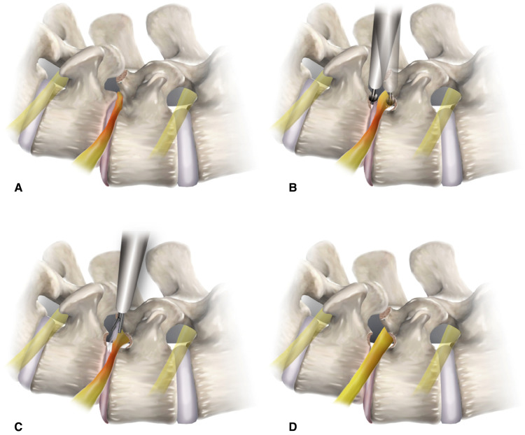
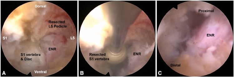
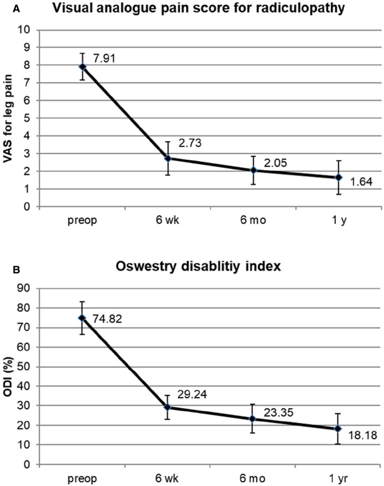
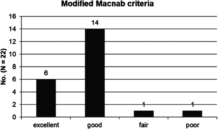
Methods: Consecutive 22 patients who suffered from radiculopathy with spondylolisthesis underwent ELF. The inclusion criterion was unilateral radicular leg pain due to foraminal stenosis in stable spondylolisthesis. After the percutaneous transforaminal approach, foraminal decompression was performed using various surgical devices under endoscopic visualization. Surgical outcomes were measured using the visual analog pain score, Oswestry disability index, and modified MacNab criteria.

Results: Pain scores and functional outcomes improved significantly during the 12-month follow-up periods. The rate of clinical improvement was 95.5% (21 of 22 patients). One patient experienced a dural tear and subsequent open surgery.

Conclusion: ELF can be effective in foraminal stenosis in stable spondylolisthesis. Technical points specializing in foraminal decompression in spondylolisthesis are required for clinical success.

Keywords: endoscopic; foraminal stenosis; foraminoplasty; foraminotomy; lumbar; percutaneous; spondylolisthesis.


Conflict of interest statement

The authors declare that the research was conducted in the absence of any commercial or financial relationships that could be construed as a potential conflict of interest.

Figures

Figure 1
Figure 1
Conceptual illustrations depicting the surgical procedure of endoscopic lumbar foraminotomy for spondylolisthesis. (A) Foraminal docking of the working sheath viewing the foraminal surgical field protecting the exiting nerve root (outside-in approach). (B) Foraminal unroofing using endoscopic burrs for resecting the upper pedicle and lower vertebral endplate. (C) Soft tissue decompression with removal of the ligamentum flavum. (D) Final point of the full-scale foraminal decompression from the axillary side to the lateral exit zone.
Figure 2
Figure 2
Intraoperative endoscopic views. Foraminal unroofing with the removal of the upper pedicle (A) and lower vertebral endplate (B) compressing the exiting nerve root (ENR). After the full-scale decompression, the ENR was freely released from the proximal axillary zone to the lateral exit zone (C).
Figure 3
Figure 3
An illustrative case of a 62-year-old Male patient. (A) Preoperative computed tomography (CT) images showing foraminal stenosis with spondylolisthesis at the L4-5 level. (B) Postoperative CT images showing foraminal decompression with resection of a part of the upper pedicle (arrow) and lower vertebral endplate (arrowheads).
Figure 4
Figure 4
An illustrative case of a 75-year-old Male patient. (A) Preoperative magnetic resonance image (MRI) showing foraminal stenosis with spondylolisthesis at the L5-S1 level (arrow). (B) Postoperative MRI showing foraminal decompression with removal of the protruded disc and surrounding bony tissues (arrowheads).
Figure 5
Figure 5
Clinical outcomes. (A) Visual analog pain score for radicular leg pain preoperatively and at 6 weeks, 6 months, and 1 year after surgery. (B) Oswestry disability index scores preoperatively and at 6 weeks, 6 months, and 1 year after surgery.
Figure 6
Figure 6
The global outcome according to the modified macNab criteria: excellent in 6 patients (27. 3%), good in 14 (63.6%), fair in 1 (4.5%), and poor in 1 (4.5%). Therefore, the success rate was 90.9%, and the clinical improvement rate was 95.5%.

References

    1. Knight MT, Vajda A, Jakab GV, Awan S. Endoscopic laser foraminoplasty on the lumbar spine–early experience. Minim Invasive Neurosurg. (1998) 41:5–9. 10.1055/s-2008-1052006 - DOI - PubMed
    1. Ahn Y, Lee SH, Park WM, Lee HY. Posterolateral percutaneous endoscopic lumbar foraminotomy for L5-S1 foraminal or lateral exit zone stenosis. Technical note. J Neurosurg. (2003) 99(3 Suppl):320–3. 10.3171/spi.2003.99.3.0320 - DOI - PubMed
    1. Yeung AT. The evolution and advancement of endoscopic foraminal surgery: one surgeon's Experience incorporating adjunctive techologies. SAS J. (2007) 1:108–17. 10.1016/SASJ-2006-0014-RR - DOI - PMC - PubMed
    1. Ahn Y, Oh HK, Kim H, Lee SH, Lee HN. Percutaneous endoscopic lumbar foraminotomy: an advanced surgical technique and clinical outcomes. Neurosurgery. (2014) 75:124–33. 10.1227/NEU.0000000000000361 - DOI - PMC - PubMed
    1. Knight M, Goswami A. Management of isthmic spondylolisthesis with posterolateral endoscopic foraminal decompression. Spine (Phila Pa 1976) (2003) 28:573–81. 10.1097/01.BRS.0000050400.16499.ED - DOI - PubMed

LinkOut - more resources