Differences in cytokines expression between Vero cells and IPEC-J2 cells infected with porcine epidemic diarrhea virus
- PMID: 36439802
- PMCID: PMC9686284
- DOI: 10.3389/fmicb.2022.1002349
Differences in cytokines expression between Vero cells and IPEC-J2 cells infected with porcine epidemic diarrhea virus
Abstract
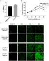
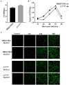
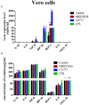
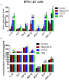
Porcine epidemic diarrhea virus (PEDV) primarily infects suckling piglets and causes severe economic losses to the swine industry. Cytokines, as part of the innate immune response, are important in PEDV infection. The cytokines secreted by cell infection models in vitro might reflect true response to viral infection of target cells in vivo. Vero cells and IPEC-J2 are commonly used as an in vitro model to investigate PEDV infection. However, it is not clear which type of cells is more beneficial to the study of PEDV. In our study, firstly, Vero cells and IPEC-J2 were successfully infected with PEDV virulent strains (HBQY2016) and attenuated vaccine strains (CV777) and were capable of supporting virus replication and progeny release. Moreover, cytokine differences expression by Vero cells and IPEC-J2 cells infected with two PEDV strains were analyzed. Compared with IPEC-J2 cells, only the mRNA levels of TGF-β, MIP-1β and MCP-1 were detected in Vero cells. ELISA assay indicated that compared to the control group, the PEDV-infected group had significantly induced expression levels of IL-1β, MIP-1β, MCP-1, IL-8, and CXCL10 in IPEC-J2 cells, while only secretion level of IL-1β, MIP-1β and IL-8 in Vero cells were higher in PEDV infected group. Finally, cytokines change of piglets infected PEDV-HBQY2016 strains were detected by cDNA microarray, and similar to those of IPEC-J2 cells infected PEDV. Collectively, these data determined that the IPEC-J2 could be more suitable used as a cell model for studying PEDV infection in vitro compared with Vero cells, based on the close approximation of cytokine expression profile to in vivo target cells.
Keywords: IPEC-J2 cells; PEDV; Vero cells; cytokines; difference.
Copyright © 2022 Yuan, Sun, Chen, Li, Yao, Wang, Guo, Li and Song.
Conflict of interest statement
The authors declare that the research was conducted in the absence of any commercial or financial relationships that could be construed as a potential conflict of interest.
Figures


Similar articles
-
Chemokines induced by PEDV infection and chemotactic effects on monocyte, T and B cells.Vet Microbiol. 2022 Dec;275:109599. doi: 10.1016/j.vetmic.2022.109599. Epub 2022 Nov 2. Vet Microbiol. 2022. PMID: 36335842
-
Comparative transcriptomic analysis of porcine epidemic diarrhea virus epidemic and classical strains in IPEC-J2 cells.Vet Microbiol. 2022 Oct;273:109540. doi: 10.1016/j.vetmic.2022.109540. Epub 2022 Aug 8. Vet Microbiol. 2022. PMID: 35987184
-
IFN-lambda preferably inhibits PEDV infection of porcine intestinal epithelial cells compared with IFN-alpha.Antiviral Res. 2017 Apr;140:76-82. doi: 10.1016/j.antiviral.2017.01.012. Epub 2017 Jan 19. Antiviral Res. 2017. PMID: 28109912 Free PMC article.
-
Transcriptome analysis of host response to porcine epidemic diarrhea virus nsp15 in IPEC-J2 cells.Microb Pathog. 2022 Jan;162:105195. doi: 10.1016/j.micpath.2021.105195. Epub 2021 Sep 24. Microb Pathog. 2022. PMID: 34571150
-
Alpiniae oxyphyllae fructus polysaccharide 3 inhibits porcine epidemic diarrhea virus entry into IPEC-J2 cells.Res Vet Sci. 2022 Dec 20;152:434-441. doi: 10.1016/j.rvsc.2022.09.011. Epub 2022 Sep 15. Res Vet Sci. 2022. PMID: 36126510
Cited by
-
Integrating network pharmacology with pharmacological research to elucidate the mechanism of modified Gegen Qinlian Decoction in treating porcine epidemic diarrhea.Sci Rep. 2024 Aug 15;14(1):18929. doi: 10.1038/s41598-024-70059-5. Sci Rep. 2024. PMID: 39147857 Free PMC article.
-
Remdesivir inhibits Porcine epidemic diarrhea virus infection in vitro.Heliyon. 2023 Nov 2;9(11):e21468. doi: 10.1016/j.heliyon.2023.e21468. eCollection 2023 Nov. Heliyon. 2023. PMID: 38027806 Free PMC article.
-
The role of innate immune responses against two strains of PEDV (S INDEL and non-S INDEL) in newborn and weaned piglets inoculated by combined orogastric and intranasal routes.Front Immunol. 2025 Jun 16;16:1584785. doi: 10.3389/fimmu.2025.1584785. eCollection 2025. Front Immunol. 2025. PMID: 40589734 Free PMC article.
References
-
- Arce C., Ramirez-Boo M., Lucena C., Garrido J. J. (2010). Innate immune activation of swine intestinal epithelial cell lines (IPEC-J2 and IPI-2I) in response to LPS from salmonella typhimurium. Comp. Immunol. Microbiol. Infect. Dis. 33, 161–174. doi: 10.1016/j.cimid.2008.08.003, PMID: - DOI - PubMed
LinkOut - more resources
Full Text Sources
Miscellaneous

